Playlist
Show Playlist
Hide Playlist
Deep and Outcropping Layer (PC) – Anatomy of the Forearm

-
Slides 06 UpperLimbAnatomy Pickering.pdf
-
Download Lecture Overview
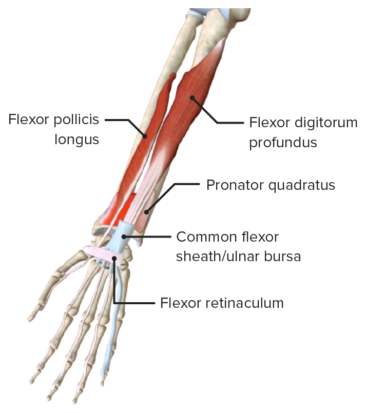
00:00 the wrist. And extensor carpi ulnaris can work with flexor carpi ulnaris to adduct the wrist. 00:00 If we now move on to some deep muscles on the posterior compartment, also known as these outcropping muscles, then we can see we have a whole series of them; abductor pollicis longus, extensor pollicis longus, extensor pollicis brevis, and extensor indicis. 00:20 Now these muscles are passing specifically to the thumb, the pollicis muscles, and to the index finger, extensor indicis. So these are passing from the forearm going across the wrist and attaching to features on digits 1 and 2. So if we look at abductor pollicis longus, abductor pollicis longus, we can see here, is passing all the way to the metacarpal of the first digit. It’s coming from the posterior surface of the radius and the ulna and also the interosseous membrane. So it has a widespread origin, and it inserts at the metacarpal of the first digit. Extensor pollicis longus, specifically, passes from the posterior surface of the ulna, and also the interosseous membrane, so not the radius this time. And it passes to the dorsal surface of the distal phalanx of the first digit; so the distal phalanx of your thumb. Extensor pollicis brevis, the posterior surface of the radius, the interosseous membrane, and it passes to the dorsal surface of the proximal phalanx of the first digit. So here we have extensor pollicis longus passing to the distal phalanx, and extensor pollicis brevis passing to the proximal phalanx. And this is where they get their names, longus and brevis. Longus, the longer of the muscle, passes most distally to the distal phalanx. And brevis, the shorter of the muscles, passing to the proximal phalanx. 01:59 All of these muscles are supplied by the posterior interosseous nerve. This originates from the deep radial nerve. And we can see they have quite a widespread function. Abductor pollicis longus extends the wrist because it crosses the wrist joint, and it works on the thumb. 02:17 It abducts the thumb and extends it at the carpometacarpal joint. Extensor pollicis longus and extensor pollicis brevis are going to work to extend the wrist, extend the distal and proximal phalanx of the thumb at the interphalangeal joints, and also extend the metacarpophalangeal and carpometacarpal joints. So we can see we have widespread function of these muscles. 02:48 Here, we can also see that we have what’s known as supinator muscle we can see here. 02:57 And supinator muscles, as its names suggest, is going to supinate the forearm. We can see that supinator is running from the lateral epicondyle of the humerus. We can see here it’s one of the lateral epicondyle of the humerus. It’s also running from the supinator fossa on the proximal ulna, so a specific region on the ulna and it passes to the posterior, lateral, and anterior surfaces of the proximal radius. Supplied by the deep branch of the radial nerve, and as its name suggest, it’s going to supinate the forearm. Finally, we can just see extensor indicis. Extensor indicis is important as it passes specifically to the index finger, given the index finger, some specific functionality. Extensor indicis is coming from the posterior surface of the ulna and also the interosseous membrane, and it passes to the extensor expansion specifically of the second digit, your index finger. It’s supplied by the posterior interosseous nerve. Again, this is coming from the deep radial nerve. 04:04 And it serves to extend the wrist and extend the second digit. You can do this independently from extensor digitorum. So the index finger is allowed to have some independent function. 04:19 So here we can see the anatomy of the posterior aspect of the forearm. We can see this is a right forearm, and we have a lot of the muscles all put together in their proper anatomical position. So we can see most distally, a whole series of tendons, and these tendons are coming from the muscles on the posterior surface. And here, we have the extensor retinaculum. 04:45 So most distally, we have the extensor retinaculum, and that prevents bowstringing when the hand is extended. When the hand is fully extended, you don’t want these tendons from splaying either side, and the extensor retinaculum prevents that. Divided into superficial and deep layers, the muscles in the posterior compartment, the tendons pass over the wrist and they’re covered in what are known as synovial sheaths. These synovial sheaths prevent friction as the tendons run over the wrist, and they create these osseous tunnels formed by the extensor retinaculum, and they help to prevent friction. So you have these synovial sheaths that prevent excess friction as the tendons are trapped between the bones of the wrist and the extensor retinaculum. And these synovial sheaths prevent that excess friction. 05:47 The tendons of the extensor digitorum flatten, as I mentioned previously, and form these extensor expansions. These are tendinous aponeurosis that pass over the dorsum and sides of metacarpal and the proximal phalanx of each digit. So instead of attaching directly to a phalanx, a distal, a middle, or a proximal, they blend with this tendinous aponeurosis that lies on the dorsum and the sides of the metacarpals and proximal phalanges. So the tendons actually just blend with this aponeurosis. It means that you actually lose extensor function. 06:31 Unlike the flexor compartment where the muscles and the tendons inserted directly onto bony points, you had a lot more flexion control. For extension, you don’t have that level of control because of this widespread insertion into the extensor expansions. So if we dig a bit deeper and remove some of these middle and superficial layers, we can actually see the deeper muscles. Here, we can see the tendons of extensor digitorum have been cut away. 07:04 So we can look into this deep aspect. We can see abductor pollicis longus here. We can see extensor pollicis longus, extensor indicis. We can see these outcropping muscles as well. 07:16 Extensor pollicis brevis is one of them. And these are passing out going across the wrist and heading towards the thumb, and for extensor indicis, the index finger. Now, this forms a very specific region at the wrist, known as the anatomical snuff box. So passing laterally towards digits 1 and 2 are these outcropping muscles. They come from deep and they pass outwards, out onto this lateral aspect heading towards digits 1 and 2, and here are those muscles that I mentioned. The tendons of extensor pollicis longus, the tendons of extensor pollicis longus, medially, and extensor pollicis brevis and abductor pollicis longus laterally, form the anatomical snuff box. So here, we’ve got the fifth digit. So this is medially. 08:23 We can see we have extensor pollicis longus. So if we follow this all the way around, we can see that we’re forming this medial boundary here. If we then look laterally, we can see we have two tendons. We have extensor pollicis brevis and we have the tendon of abductor pollicis longus. And here, we can see a small little region, this triangular region known as the anatomical snuff box. It’s located on the lateral surface of the wrist, bound by these tendons. You can also observe that the index finger receives a specific muscle and this is where it separates and distinct to extensor digotorum, and this is extensor indicis. We'll come back to the anatomical snuff box when we look at the dorsum of the hand. So if we then look at the posterior compartment, we can appreciate some important nerves which are located here. 09:24 Most of these arteries are going to be a direct continuation of the brachial artery. So here, we can see we have various tributaries running along the posterior compartment. So we can see the posterior interosseous artery, and that’s coming from the ulnar artery. 09:43 And then we can see here, we’ve got the radial nerve that’s passing into the posterior compartment to supply all of these muscles. They’re running in the posterior compartment. 09:57 So in this lecture, we’ve looked at the forearm in cross-section. We’ve looked at the antebrachial fascia and the compartments that it forms. We then looked at a whole series of muscles in the anterior compartment, and we looked at their relations to one another within the anterior compartment. And then we looked at the posterior compartment. 10:15 We looked at the extensor muscles, where again, we looked at a whole series of muscles. And then briefly at the end, we looked at the neurovascular relations. We’ll look at the neurovascular relations in more detail when we look at the overview of the blood supply to the upper limb as a whole, and that’s in a later lecture.
About the Lecture
The lecture Deep and Outcropping Layer (PC) – Anatomy of the Forearm by James Pickering, PhD is from the course Upper Limb Anatomy [Archive].
Included Quiz Questions
Which statement concerning the cubital fossa is correct?
- The brachioradialis forms its lateral border.
- It is a diamond-shaped region anterior to the elbow joint.
- Its floor is formed by the pronator teres muscle.
- The flexor carpi radialis forms its medial border.
- It has the ulnar nerve coursing through it.
Which bony landmark is associated with the common flexor origin?
- Anterior surface of the medial epicondyle
- Anterior surface of the lateral epicondyle
- Posterior surface of the lateral epicondyle
- Posterior surface of the medial epicondyle
- Lateral surface of the medial epicondyle
Which bony landmark is associated with the common extensor origin?
- Posterior surface of the lateral epicondyle
- Anterior surface of the lateral epicondyle
- Anterior surface of the medial epicondyle
- Posterior surface of the medial epicondyle
- Medial surface of the medial epicondyle
Which statement concerning the ulnar nerve is correct?
- It supplies the flexor carpi ulnaris.
- It enters the hand by passing through the carpal tunnel.
- It courses around the lateral epicondyle.
- It is derived from the lateral cord of the brachial plexus.
- It supplies none of the flexor digitorum profundus.
Which statement concerning the ulnar nerve is correct?
- If it is severely compressed at the level of the elbow or upper arm, it leads to what is termed the claw hand.
- It courses through the cubital fossa lateral to the brachial artery.
- It courses between the flexor digitorum profundus and the interosseous membrane in the anterior compartment of the forearm.
- It is derived from the posterior cord of the brachial plexus.
- It supplies all the muscles in the anterior compartment of the forearm.
To which bony structures do the tendons of the flexor digitorum profundus attach?
- The distal phalanges of the medial 4 fingers
- The proximal phalanges of the medial 4 fingers
- The middle phalanges of the medial 4 fingers
- The heads of the medial 4 metacarpals
- The bases of the medial 4 metacarpals
Which statement concerning the brachioradialis is correct?
- It is involved in returning the forearm to a mid-prone position (tennis racket grip).
- It is supplied by the median nerve.
- It forms the medial (ulnar) boundary of the cubital fossa.
- It originates from the lateral epicondyle of the humerus.
- It can flex the hand at the wrist joint.
Author of lecture Deep and Outcropping Layer (PC) – Anatomy of the Forearm

James Pickering, PhD
Customer reviews
5,0 of 5 stars
| 5 Stars |
|
5 |
| 4 Stars |
|
0 |
| 3 Stars |
|
0 |
| 2 Stars |
|
0 |
| 1 Star |
|
0 |
