Playlist
Show Playlist
Hide Playlist
Cranial Nerve V: Trigeminal nerve

-
Slides 7 CranialNerves 1 BrainAndNervousSystem.pdf
-
Reference List Anatomy.pdf
-
Download Lecture Overview
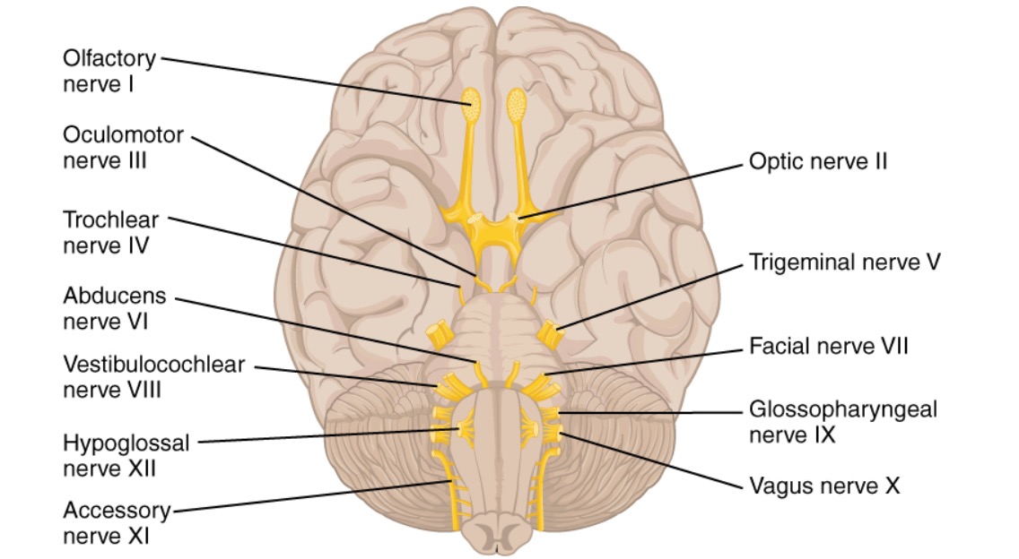
00:00 The trigeminal nerve, cranial nerve number V is a complex nerve. It has three divisions or nerves associated with it. The anatomic components of the trigeminal would be the ophthalmic nerve shown here as V/1. This is transmitted through superior orbital fissure. 00:22 Then there is the maxillary nerve as the second division or component of your trigeminal nerve, V/2. 00:29 This division is transmitted through the foramen rotundum. Your third and final anatomic component of your trigeminal nerve is the mandibular nerve. 00:37 It is transmitted through the foramen ovale and it is termed here as V/3. 00:45 Functional components of the trigeminal, there are two. One functional component is general somatic afferent. Then because the trigeminal nerve innervates the first pharyngeal arch, it also has branchial efferent functional components. If we look at the trigeminal nerve with respect to general somatic afferent connections or information as being conveyed and do so by each division or nerve, we’ll start with the ophthalmic division, V/1. That area is shown here in yellow. The ophthalmic nerve will convey sensory information from the eyes in conjunctiva. The upper eyelid which is in this shaded area, orbital contents are within the shaded area as well. What we cannot see in this particular image is that sensory information from the frontal sinuses and ethmoidal air cells is being conveyed by this nerve. The dorsum of the nose which we can see right along here, anterior scalp shown in through here and then the dura mater in the anterior cranial fossa as well as a reflection of the dura which would include a portion of the tentorium cerebelli. With respect to the maxillary division, general somatic afferent information being conveyed by V/2 is shown here in this region. This is conveying sensory information from the nasopharynx and nasal cavity, the palate. Maxillary teeth are within this shaded area, the maxillary sinus, skin from the lower eyelid in through here down to the upper lip. 03:05 This nerve will also innervate dura mater and it’s the dura found in the middle cranial fossa. 03:13 V/3, the mandibular division conveys general somatic afferent information from the skin of the lower lip and then just below that. We can see that area here in green, the anterior external ear and a portion of the external acoustic meatus, the temporal fossa, as well as the anterior two-thirds of the tongue for general somatic information. In addition, general somatic information is conveyed from the mandibular teeth since they’re within this shaded area. The air cells that we find in the mastoid process, mucous membranes of the cheeks, mandibular area, and then these two will also have innervation from the dura mater in the middle cranial fossa. The branchial distribution from the trigeminal, those branchial efferents is going to be conveyed through the mandibular division, V/3. The muscles that are derived then from the first pharyngeal arch are going to be the muscles of mastication. We have four pairs: the temporalis muscle, the masseter, lateral pterygoid, and then lastly for you to remember will be the medial pterygoid. 05:01 In addition to those muscles of mastication, additional branchial efferents are going to innervate the tensor tympani, the tensor veli palatini, anterior belly of digastric, and then we also have innervation of the mylohyoid through these branchial efferents. A clinical consideration for the trigeminal nerve is shown here. 05:34 This is demonstrating herpes zoster ophthalmicus. You can see the blisters here of the herpes virus in the ophthalmic distribution. This is an emergent condition. It’s associated with pain. It’s an emergent condition because blindness is indeed a risk in this condition. You can see in this inflammatory state, you can see how red the conjunctiva is. Another clinical consideration that involves the trigeminal nerve is that of trigeminal neuralgia. This is also referred to as tic douloureux. The symptoms of tic douloureux are going to be pain in one or more regions supplied by the divisions of the nerves, so again we have ophthalmic, maxillary, mandibular. The ophthalmic division is rarely involved in tic douloureux. 06:45 It’s more common in either the maxillary or mandibular distributions. A fairly common cause of this is that there’s compression of the trigeminal nerve itself by either the superior cerebellar artery which is shown here and that comes close to actually involving the trigeminal nerve. If it gets too close and it’s pulsating on the trigeminal nerve, over time, those micropulsations will cause demyelination. 07:24 Then the nerve fibers lose their insulation and then they start to have cross-talk between one another. That conveys pain back to the central nervous system. It could be due by compression from another neighboring artery as well but the superior cerebellar artery is a common culprit in this process. 07:46 A surgical procedure can decompress or alleviate that compression. Tumors could also cause tic douloureux as can aneurysms and infarctions are also a possible cause of this clinical condition.
About the Lecture
The lecture Cranial Nerve V: Trigeminal nerve by Craig Canby, PhD is from the course 12 Cranial Nerves and Their Functions.
Included Quiz Questions
Which of the following statements regarding the trigeminal nerve is NOT true?
- The maxillary division of the trigeminal nerve is transmitted through the superior orbital fissure.
- It has three divisions.
- It innervates the muscles of mastication.
- One of the functional components of the trigeminal nerve is the branchial efferent.
- It is a mixed (sensory and motor) cranial nerve.
Mandibular branch of the trigeminal nerve carries which of the following sensations from the anterior two-thirds of the tongue?
- Pain sensation
- Protrusion of the tongue
- Gustatory sensation
- Depression of the tongue
- Retraction of the tongue
Which of the following muscles is not innervated by the trigeminal nerve?
- Posterior belly of the digastric muscle
- Masseter muscle
- Temporalis muscle
- Medial pterygoid muscle
- Lateral pterygoid muscle
Which statement about tic douloureux is correct?
- The compression of the trigeminal nerve by the superior cerebellar artery is a possible cause.
- It presents as continuous, stabbing facial pain.
- Patients cannot precisely localize the pain.
- Maxillary and mandibular subdivisions of the trigeminal nerve are rarely involved.
- It occurs most commonly in the ophthalmic subdivision of the trigeminal nerve.
Author of lecture Cranial Nerve V: Trigeminal nerve

Craig Canby, PhD
Customer reviews
5,0 of 5 stars
| 5 Stars |
|
1 |
| 4 Stars |
|
0 |
| 3 Stars |
|
0 |
| 2 Stars |
|
0 |
| 1 Star |
|
0 |
I do like it very much its straight forward . He has shown all the main points . Many thanks indeed.
