Playlist
Show Playlist
Hide Playlist
Cranial Fossa and Nerves

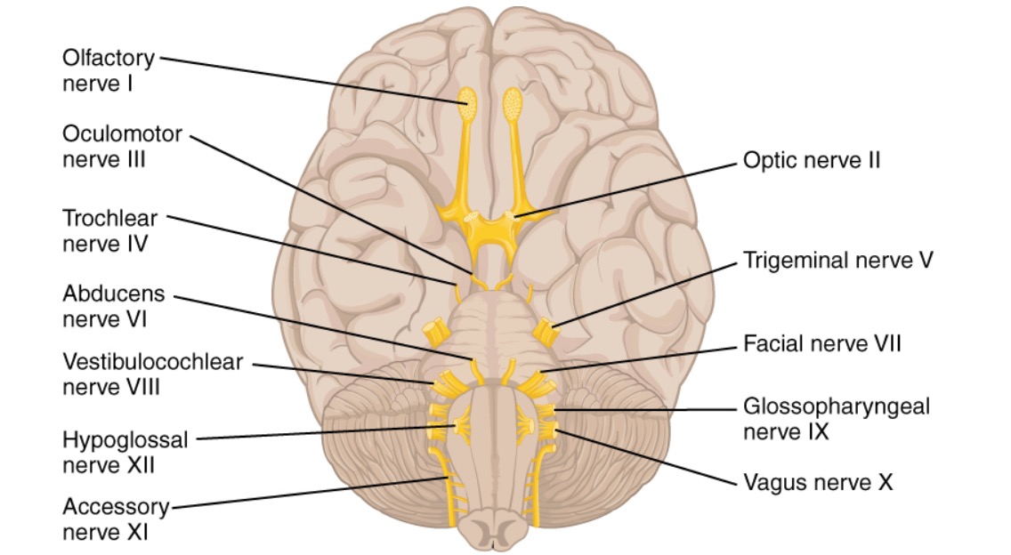
00:01 Okay, that’s about lymph nodes. Moving up into the face. To understand the anatomy of the face at your level, you need to have a bit of understanding about the cranial fossa, as well as the foramen. That leads to your cranial nerves. So, your cranial fossa has got an anterior cranial fossa at the front, middle cranial fossa and posterior cranial fossa. What is the first cranial nerve? Olfactory. 00:38 Olfactory. What’s the function, action of olfactory? Smell. Smell. Where does it, how does it come in to the nose for the function? Starts from the skull base through the cribriform. 00:52 Yeah, it sends fibers through the cribriform plate. It doesn’t have a foramen, it comes through the cribriform plate. So that’s why when you have base of skull fracture, you have CSF leak coming through the nose because of fracture of cribriform plate. So the olfactory, it just got those sensory fibers into the nose coming through the cribriform plate. Okay, that’s all you need to know. Second is optic. 01:17 Optic comes out through? Optic. Canal. 01:22 Optic canal. Does it accompany anything else? Ophthalmic artery. Ophthalmic artery, very good. Where does ophthalmic artery come from? First branch of internal carotid. 01:32 Very good. Tell me, branch of the? It’s a branch of internal carotid. So there’s your ophthalmic, coming out through optic canal, accompanied with ophthalmic artery. 01:45 What’s next? Oculomotor. So tell me how oculomotor comes out? Superior orbital fissure. Very good. Okay, tell me what are the other structures in the superior orbital fissure. Cranial nerves III, IV, VI, V1, emissary veins, I don’t know. You’re right, pretty much, you got all the important ones. And then you have the superior and inferior ophthalmic veins which for the purpose of your part you don’t need to worry too much but you need know three, that’s your occulomotor, abducens and trochlear. So III, IV, and VI cranial nerves as well as the first division of the trigeminal comes out through the superior orbital fissure here, okay? So III, IV, first part of V and VI. 02:45 Trigeminal nerve has got three components, ophthalmic, maxillary, mandibular. So ophthalmic is the one that you said comes out of the superior orbital fissure. So when you are anesthetizing somebody’s forehead to excise a lipoma here, what nerve are you anesthetizing? It has to be from the ophthalmic division of trigeminal. So it is normally your supraorbital and the supratrochlear nerves, because they are the ones you are anesthetizing when you are doing anything in the forehead, okay? You also have lacrimal gland branches, ethmoidal, but you don’t have to go into detail. All you need to remember for this exam is superior orbital fissure, what are the nerves that come out. We have covered I, II, III, IV, V, VI. Okay what’s next? Seventh cranial nerve, the facial nerve. How does it come out? It comes, I think through the internal acoustic meatus? Internal acoustic meatus. 03:46 Stylomastoid foramen. And there’s a facial canal? It’s the facial canal. Yup! Well then, it gives off a tympanic branch and then it sends through the stylomastoid foramen branch off into five branches. Okay, that’s very good. I think we will come to the facial nerve branches when we do the face, but at this point you need to remember how does it come out. So facial nerve comes out the internal acoustic meatus along with the 8th cranial nerve, with the vestibulocochlear. So VII and VIII comes out of the internal acoustic meatus. The VIII cranial nerve stops at the middle ear, it doesn’t come out further than that. VII cranial nerve, which is the facial, come enters the facial canal, then comes out the stylomastoid foramen and then it comes to the face. 04:37 It gives a few branches inside. One is your nerve to stapedius, anything else? This is quite important. Facial nerve is quite an important nerve. So you need to know a bit more about the facial nerve. Now I’m sure all of you know this, the branches, that’s after it comes out from stylomastoid foramen but before that, think about it. Nerve to stapedius, one, second is the chorda tympani. Chorda tympani. So what does this chorda tympani do, apart from taste to the -- Taste. Taste and something else. 05:14 Soft palate sensation? Soft palate and tonsils. Yes, you can get sensation to the palate as well, but anything to do with submandibular gland? That’s right. It sends parasympathetic fibers to the submandibular gland as well, so that’s part of the facial nerve coming through the chorda tympani. Any other branch? So nerve to stapedius, chorda tympani and one more important branch, greater petrosal nerve, okay? Greater petrosal nerve. What does that do? Greater petrosal nerve predominantly carries preganglionic parasympathetic fibers. 06:03 Yes, to the lacrimal gland, to the lateral side of the nose, to the ethmoidal region here. So, that’s actually when you get hay fever, your tears, runny nose, it’s all due to the greater petrosal nerve, which is a branch of the facial, okay? Right, VIII cranial nerve we discussed vestibulocochlear. 06:30 What’s IX? Glossopharyngeal. How does it come out? From the -- No. 06:37 Is it the jugular canal? Jugular foramen. 06:41 Foramen, sorry. Okay, what other nerves coming out of jugular foramen? X. 06:45 IX, X, XI, okay? IX, X, XI cranial nerves come out through the jugular foramen. 06:53 Then of course you have the internal jugular vein going in, and is also accompanied by the ascending pharyngeal artery, okay? So, jugular foramen is quite big, quite important. So three cranial nerves coming out, the jugular vein going in, along with the ascending pharyngeal artery. 07:15 So that’s IX, X, XI. XII, hypoglossal nerve comes out through the hypoglossal canal. 07:20 Question, should we also learn the cavernous sinus as well, what is that? 3rd, 4th and yeah. So these are the main nerves.
About the Lecture
The lecture Cranial Fossa and Nerves by Stuart Enoch, PhD is from the course Head and Neck Anatomy—MRCS.
Author of lecture Cranial Fossa and Nerves

Stuart Enoch, PhD
Customer reviews
5,0 of 5 stars
| 5 Stars |
|
5 |
| 4 Stars |
|
0 |
| 3 Stars |
|
0 |
| 2 Stars |
|
0 |
| 1 Star |
|
0 |
