Playlist
Show Playlist
Hide Playlist
Ciliary Body and Processes
-
Slides 16 Human Organ Systems Meyer.pdf
-
Reference List Histology.pdf
-
Download Lecture Overview
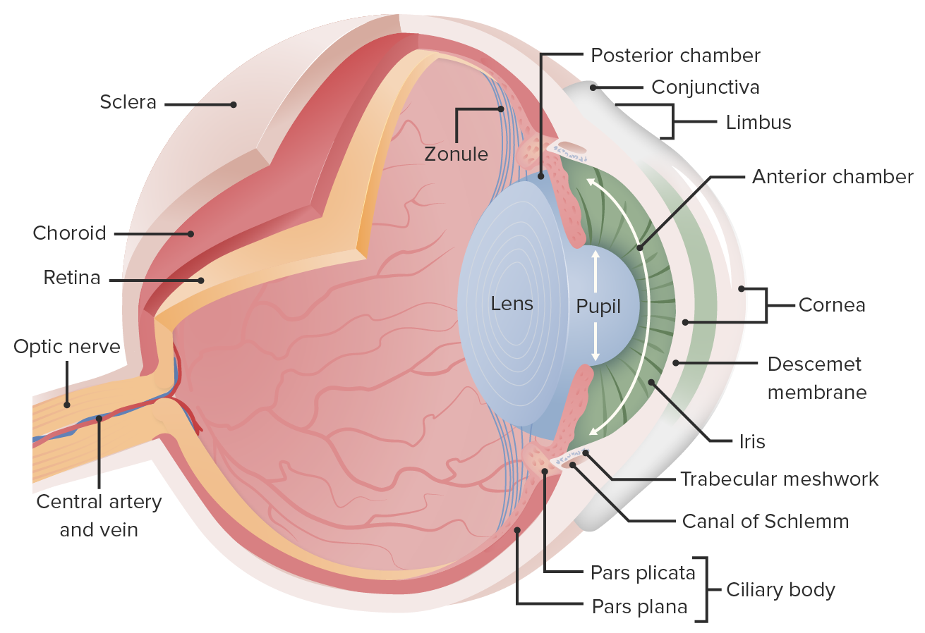
00:01 Let me now move from the iris to the ciliary body shown here in the diagram. 00:06 It has got a number of components, which I would like to explain to you. First of all the ciliary muscle is the bulk of what you see on the top left-hand side of this picture. Ciliary muscle is very important because when it contracts or relaxes, it affects the dimensions of the lens because the ciliary processes which you see there, they have an epithelium on them. There's two epithelial layers there and again the extensions on the neural part of the retina. Those epithelial cells closest to the posterior chamber or the vitreous actually make these little fibres and those fibres extend to the lens. And as we will see in a moment when the ciliary muscles contract or relax, that changes of tension on those fibres extending into the lens and, therefore, the shape of the lens. So the ciliary process is very important for that and also the epithelial cells produce the aqueous humor that I will explain in a moment. So let's just go back and look at the image on the right-hand side. Let us look very carefully. You can see some of these zones with fibres extending from the ciliary processes. They are very hard to see in sections because often when you section through the eye, it does not have to be lucky enough to get the zonular fibres, but some are shown here if you look very very finely and top of the lens are shown up in the top left hand side of this particular image. And there is a ciliary muscle there in the corner and on the right-hand side of that, the dark green structure is going to be the sclera. You are looking right at the junction between the cornea and the sclera in an area we call the limbus. Let us just go back a step before we continue with those zonular fibres and the lens and have a closer look at this double epithelium that lines the ciliary processes. You can see two epithelia here adjacent or facing the posterior chamber or the vitreous is the non-pigmented ciliary epithelial cells, the cuboidal cells and then below them are the pigmented ciliary epithelial cells and then below those cells is going to be the stroma of the ciliary body. And again let me remind you that this double epithelial layer is a nonneural extension of the retina. Back in the posterior part of the retinal course, there are going to the photoreceptor cells and also this pigmented epithelium. Right between the two cell layers is the ciliary channel, which is an important collection site when the process of these cells are making the aqueous humor. And if you look very very carefully at the top part of the epithelium, the nonpigment epithelium, you can see a very fine line running across the surface of those cells. That happens to be the basal lamina, the basement membrane of this epithelial layer. The basement epithelium of the pigmented epithelium is adjacent to the stroma. 03:41 So really that ciliary channel you see labelled there has both the apical surfaces of the non-pigmented epithelium and also the apical surfaces of the pigmented epithelial cells. And we are going to see in a moment that basal lamina on the surface, on the non-pigmented layer are going to be the component that anchors the zonula fibres that extend then to equator of the lens. 04:14 Here is a diagram explaining that double epithelium that I indicated to you in the previous slide. I actually like this diagram. I like the colour in that. 04:27 So congratulations to the artist. Let us focus on the bottom part of the diagram, you can see a fenestrated capillary. That is the stroma component of the ciliary processes in the ciliary body and just above that is the pigmented epithelial cell. Then the ciliary channel and then the non-pigmented epithelial cells. And then you can see the basal lamina on the non-pigmented ciliary epithelial cells and extending from that basal lamina are the zonula fibres that are going to go the equator of the lens. And then above that it just tells you really the components of the aqueous humor. They are produced, those components, by the epithelial cells particularly the non-pigmented cells. They are very very busy. 05:25 They are very easily processing various components coming up from those fenestrated capillaries. They're actively processing materials. So they have lots of sodium and potassium pumps etc and a very busy epithelium that makes up this layer. Busy because not only producing the aqueous humor, but they are also producing those zonula fibres as well. 05:54 So they are busy cells. And that aqueous humor is going to circulate through the anterior and posterior chambers of the eye. Now I can show you the canal of Schlemm which sits in the limbus that I pointed out earlier. There is a corneal epithelium coming around to join the conjunctiva and the cornea is continuous with the sclera and there's the canal of Schlemm on the left hand side in low magnification and on the right hand side in the centre at high magnification. This is the network that is going to allow that aqueous humor to drain into it and finds its way into the venous system. Here is a diagram explaining that, the pictures on the left-hand side and the diagram just follow the sequence. 06:48 One, the epithelial cells produce the aqueous humor that circulates from the posterior chamber through the little valve created by the iris into the anterior chamber and then into the canal of Schlemm and then to the venous system. That is constantly being produced and it maintains a certain pressure in the anterior and posterior chamber of the eye. If there's some reason that there is a blockage in that canal or over production, then the pressure increases and that is a condition that we call glaucoma and is quite a common eye disease that needs to be treated. The vitreous cavity shown in the very bottom part of the diagram is a gel-like component. It is a large component. Its job being a gel is to maintain the shape of the eye, but it is also a shock absorber. When you do sudden eye movements, the vitreous protects the retina the very sensitive retina from vibrations caused by those rapid eye movements. You can see the lens there and you can see the zonular fibres extending from the epithelium are described earlier to the equator of the lens. Again let me remind you that post lens fibres or zonular fibers are produced by the epithelial cells that produce the aqueous humor.
About the Lecture
The lecture Ciliary Body and Processes by Geoffrey Meyer, PhD is from the course Sensory Histology.
Included Quiz Questions
Which of the following best describes the double epithelial layer of the ciliary processes in the eye?
- It is a non-neural extension of the retina.
- The pigmented layer is in direct contact with the posterior chamber.
- The pigment has a different composition than that found in the iris.
- The non-pigmented cells are in contact with the stroma of the ciliary body.
- The zonula fibers are produced by the pigmented epithelium.
Zonular fibers originate from which of the following?
- Basal laminae of the non-pigmented epithelium of the ciliary body
- Pigmented epithelium of the ciliary body
- The lens
- Posterior lens capsule
- Canal of Hannover
What is the correct anatomical sequence of structures of the ciliary body from the aqueous to the ciliary stroma?
- Zonular fibers, basal lamina of non-pigmented epithelium, non-pigmented epithelium, pigmented epithelium, basement membrane of pigmented epithelium
- Zonular fibers, basal lamina of non-pigmented epithelium, non-pigmented epithelium, basement membrane of pigmented epithelium, pigmented epithelium
- Zonular fibers, non-pigmented epithelium, basal lamina of non-pigmented epithelium, basement membrane of pigmented epithelium, pigmented epithelium
- Zonular fibers, non-pigmented epithelium, basal lamina of non-pigmented epithelium, pigmented epithelium, basement membrane of pigmented epithelium
Customer reviews
5,0 of 5 stars
| 5 Stars |
|
5 |
| 4 Stars |
|
0 |
| 3 Stars |
|
0 |
| 2 Stars |
|
0 |
| 1 Star |
|
0 |


