Playlist
Show Playlist
Hide Playlist
Chronic Obstructive Pulmonary Disease (COPD): Tests and Investigations


-
Slides 04 COPD RespiratoryAdvanced.pdf
-
Download Lecture Overview
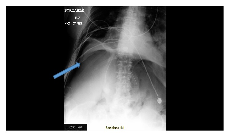
00:00 The other aspect of lung function tests that you need to think about, are the flow volume loops and the lung volumes. So, the lung volumes are increased in COPD, the patient has bigger overall lung volumes than normal. That may seem surprising but that relates to the air trapping that we discussed that I mentioned earlier, and that I'll discuss in more detail in a second. The reason for the increase in lung volume is that the residual volume, the amount of air left in the lung at the end of expiration, increases. So although the lung volumes are larger, it's useless volume, it's not air that shifting during inspiration and expiration. It's just the residual volume that's driving the increase in lung volume. If the patient has emphysema, then they've lost a lot of surface area for gas exchange because of the alveolar destruction. And that means the transfer factor which measures the ability of oxygen to get in from the lungs into the blood, goes down. So a low transfer factor suggest emphysema. 01:11 In addition, the alveolar structures are required for splinting open the airways, as you breath out. 01:18 So on expiration, there tends to be, there's a positive pressure in the thoracic cavity, and that squeezes air out of the lungs, but it also squeezes the bronchi, taking the air out of the lungs. And if the bronchi have lost their structural support, then there'll be a tendency for them to be closed off by that pressure, the expiratory pressure. 01:39 And that's called dynamic airways collapse and occurs in emphysema where the loss and destruction of the alveoli means the bronchi are not splintered open and cannot combat the positive pressure of expiration. And you can see that on a flow volume loop, which is illustrated here on the right hand side of the slide. You can see that the person with emphysema, the solid blue line, what happens is that as they breathe out there’s a rapid increase in flow initially, and then there is a sudden decrease in flow due to this dynamic airways collapse, and that’s followed by a prolonged phase of low flow expiration. The other investigations you need to do with somebody with COPD, will clear your chest X rays is useful, but actually it’s not that abnormal in most patients. In patients with significant disease, it will show a degree of hyper-inflation with reduced lung markings, more visible anterior rib ends than normal, perhaps flattened diaphragms and a small heart. You can see bullae on a chest X ray so some people who have quite expanded lung cysts due to COPD, that is bullus that is visible on a chest X ray. And also a chest X ray is important to identify complications, pneumonia, pneumothorax, and chronically, to make sure that they don’t have cancer. 02:52 The CT scan is used to identify patients who have emphysema, to look for bullae, and perhaps to look for co-existent bronchiectasis or other complications. So, these are some examples of X rays you might see. The X ray on the left hand side of the slide is somebody with a hyperexpanded lungs due to COPD and basically you see black lungs, very long thin lungs, and the heart is stretched as well, and that’s a hyperexpanded pair of lungs with oligaemic lung fields. The middle CT scan shows centrilobular emphysema, and what you can see a black hole is where there’s been lung destruction with the grey material around the outside being the normal lung. That’s a sort of Swiss cheese lung, it’s got holes in it. The last scan is somebody who also has some emphysema but that’s distributed in a different way, it’s different in being centrilobular in the middle of the lung lobules. It’s actually around the septum, around the edges of the lung lobules, around the edges of the lung. What other investigations are useful in COPD? Well, actually not much. Lung function, chest X ray, some patients need a CT scan, blood tests, you may want to measure the alpha-1-antitrypsin levels to see whether they have early onset emphysema, if they are relatively young patients, especially if they have basal emphysema on their CT scan. Normal blood tests for blood count, Fbc, U+E, LFT are all normal. Some patients, you need to do an echocardiogram and ECG and there’s two reasons for that- one, the main differential diagnosis for somebody with COPD would be cardiac breathlessness, congestive cardiac failure, or a valvular problem such as aortic cyanosis, and you may need to exclude that as a cause of their breathlessness. 04:34 And the other reason why you need to do an echocardiogram and ECGs is those patients with chronic hypoxia may develop cor pulmonale and you need an echocardiogram to measure the pulmonary hypertension that could be present in those circumstances. Invasive tests such as bronchoscopy or lung biopsy are not needed for patients with COPD, they are not needed for diagnosis at all. 04:53 So, how do you recognize patients with COPD? It’s a combination of gradually worsening breathlessness on exertion over years or months with a significant pack year history, 25 to 30 pack year history, therefore, most patients will be aged over 50 or so. Plus obstructive spirometry. 05:13 The chest X ray often looks normal, the main differential diagnosis is congestive cardiac failure, chronic PEs, that will be a shorter history, and they’ll have an abnormal transfer factor, and importantly the lung volumes and spirometry will be normal. Chronic asthma, there’s no history of smoking, there’s definitely a past history of asthma in most of those patients. And pulmonary fibrosis. And this is where crackles are important, because if you hear crackles, that’s not due to COPD, and in addition the lung function pattern you get with pulmonary fibrosis is different, it’s a restrictive lung function with an increased FEV1 to FVC ratio and usually a fall in transfer factor as well.
About the Lecture
The lecture Chronic Obstructive Pulmonary Disease (COPD): Tests and Investigations by Jeremy Brown, PhD, MRCP(UK), MBBS is from the course Airway Diseases.
Included Quiz Questions
Which of the following is NOT a feature of lung function in an emphysematous patient?
- Decreased residual volume
- Increased total lung volume
- Low transfer factor
- Dynamic airway collapse on expiration
- Dip in the expiratory curve on the flow volume loop
Which of the following is responsible for a rapid fall in expiratory flow on the flow volume loop in a patient with severe emphysema?
- Dynamic airway collapse
- Increased total lung volume
- Decreased residual volume
- Increased peak expiratory flow
- Air trapping
Which of the following is NOT a finding on the X-ray of a patient with severe emphysema?
- Boot-shaped heart
- Increased intercostal distance
- Prominent bullae
- Reduced lung markings
- Flattened diaphragm
Which of the following complications is typically responsible for pneumothorax in a patient with COPD?
- Subpleural bullae
- Hyperinflated lungs
- Expanded intercostal spaces
- None of the options
- Infections
Which of the following is NOT a common complication of emphysema?
- Cavitation
- Pneumothorax
- Bronchiectasis
- Pneumonia
- Respiratory failure
What is the purpose of periodically performing an echocardiogram on a patient with COPD?
- To monitor for pulmonary hypertension
- To monitor for arrhythmia
- To monitor for structural heart changes (e.g., tubular heart)
- To monitor for valvular defects
- To monitor for pneumopericardium
Which of the following is NOT a useful diagnostic tool for COPD?
- Bronchoscopic biopsy
- Chest X-ray
- CT scan
- Pulmonary function test
- Spirometric analysis
Author of lecture Chronic Obstructive Pulmonary Disease (COPD): Tests and Investigations

Jeremy Brown, PhD, MRCP(UK), MBBS
Customer reviews
5,0 of 5 stars
| 5 Stars |
|
5 |
| 4 Stars |
|
0 |
| 3 Stars |
|
0 |
| 2 Stars |
|
0 |
| 1 Star |
|
0 |
