Playlist
Show Playlist
Hide Playlist
Choroid and the Retina
-
Slides 16 Human Organ Systems Meyer.pdf
-
Reference List Histology.pdf
-
Download Lecture Overview
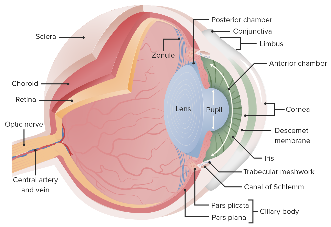
00:01 Let's now move back and look at the choroid. 00:06 I mentioned earlier it's supports the retina. 00:09 Shown in this slide, It's a very it's a pigmented layer. 00:13 It's got pigment with in the stroma, but it's dominated by capillary by blood vessels these blood vessels also pass on to the ciliary body the ciliary processes and you can see those blood vessels when you examine the internal structure of the eye with the ophthalmoscope. 00:33 That pigment and the pigmented epithelium of the retina. 00:38 Help to stop scattering of light around the inside of the eye chamber and therefore glare. 00:48 On the double pigmented layer on the posterior aspect of the IRS that absorbs light coming in. 00:55 And as I said if it's reflected back, it can determine the color of your eyes depending on the population of melanocytes in the stroma, but the choroid essentially is a vascular layer. 01:08 It's important because as I mentioned earlier in the lecture, it provides nutrition to the avascular retina, particularly the very important ganglion cells neural cells and also the photoreceptors and below you can see it's continuous dealing with the outer fibrous coat of the eye the sclera. 01:30 On this slide, you can see on the right hand side the sclera colored green and then there's choreo capillary layer. 01:38 You can just see little red blood vessels in this capillary layer, and it's separated from the pigmented epithelium and the retina by this membrane is a very thin faint membrane that runs between the pigmented epithelium and the choroid. 01:54 It forms part of the barrier the blood retinol barrier. 02:01 Well, let's look at the retina and goodness me. 02:06 Look how complicated it is. 02:09 On the right hand side is a picture taken from a light microscope of the retina and all its layers supported by the pigmented epithelium and the choroid. 02:22 And that coincides with a diagram in the center and then the picture in the center, I should say and a diagram on the left hand side. 02:30 I'm not going to go through all these cells and all the process of vision. 02:35 As I said earlier it see the role I will leave to the neurophysiologists and the ophthalmologist, but I'll just point out a couple of things. 02:44 On the diagram, focus on the rods and the cones they're shaped differently. 02:52 They have outer segments that are in contact with the retinal pigment epithelium. 03:00 Those rods and cones are in various concentrations around the retina. 03:05 Cones are specialized for color, vision, and rods for vision in dim light and peripheral vision. 03:16 There is a part of the eye the macular, and in the macula, which is about four or five millimeters in diameter. 03:26 There is a central part of that macular called the fovea centralis. 03:30 It's about one and a half maybe two millimeters in diameter. 03:37 And within that the central part of that is a foveola. 03:41 It's about 200 microns in diameter. 03:45 That's fully concentrated by cones only and that's where our focus of light rays is perceived to be the strongest. 03:57 It's where we can perceive the most detail of structures and it's where color is perceived. 04:05 It's a high concentration as a set of cones those cones are quite large. 04:10 They can be 80 to 60 microns in height, the rods a little bit smaller. 04:19 And as I said those cones are concentrated in that foveola, there's about 4,000 of them there. 04:27 And they occupy that space and also if you look at this diagram at the foveola the central spot, the retina is slightly tilted on an angle so that the light actually hits the photoreceptor cells without passing through all the layers of the ganglion cells and neural cells supporting those cells those photoreceptors. 04:53 In that area in the foveola, there are 4,000 cones as I pointed out and the ratio of cones to the ganglion cells as 1 to 1, whereas elsewhere, It's much higher than that. 05:07 There are in fact about a hundred and twenty million rods. 05:12 And only about 5 to 7 million cones. 05:16 So about a 125 to 127 million photoreceptors. 05:22 There's only a million ganglion cells. 05:25 So each gang and cell their for sends impulses from a combination of these photoreceptors. 05:34 I want to just turn your attention now to the pigment epithelium that pigment epithelium is a very important structure that has a number of different jobs to do. 05:44 The outer segments of the rods and cones consist of discs containing the visual pigment which are shared continually and one job of these retinal pigment epithelial cells is to phagocytosis. 05:59 Those discs being shed. 06:01 And then to also re photosynthesize the visual pigment that has been dissociated in contact with light. 06:11 They also form a very important blood retinol barrier. 06:17 So they're very very important cells. 06:20 And as I said the pigment helps to stop light from being reflected back into the eye and therefore creating glare.
About the Lecture
The lecture Choroid and the Retina by Geoffrey Meyer, PhD is from the course Sensory Histology.
Included Quiz Questions
Bruch’s membrane lies between which of the following structures?
- Pigmented epithelium and choroid
- Choroid and basal lamina
- Pigmented epithelium and nonpigmented epithelium
- Retina and ciliary body
- Lens capsule and epithelium
Which of the following layers lies immediately superficial to Bruch’s membrane?
- Choriocapillaris
- Sattler's layer
- Haller's layer
- Pigmented epithelium
- Basal lamina
Color blindness primarily results from a defect in which of the following?
- Cone cells
- Rod cells
- Choriocapillaris
- Bruch’s membrane
- Ganglion cells
Which of the following is the main function of the rod cells in the retina?
- Peripheral vision
- Structural integrity
- Color vision
- Provision of high visual acuity
- Prevention of nystagmus
Customer reviews
2,0 of 5 stars
| 5 Stars |
|
0 |
| 4 Stars |
|
0 |
| 3 Stars |
|
0 |
| 2 Stars |
|
1 |
| 1 Star |
|
0 |
There were good pictures and diagrams but without a pointer showing what structure he's referring the whole lecture was hard to follow. I read through the transcript too and I feel like the explanations did not really match what was being showed on the slides (he kept talking about the numbers and diameters of things which were not shown on the slides).


