Playlist
Show Playlist
Hide Playlist
Cholelithiasis vs. Choledocholithiasis with Case

-
Slides Gastroenterology 14 LBSD Gallstone Diseases.pdf
-
Reference List Gastroenterology.pdf
-
Download Lecture Overview
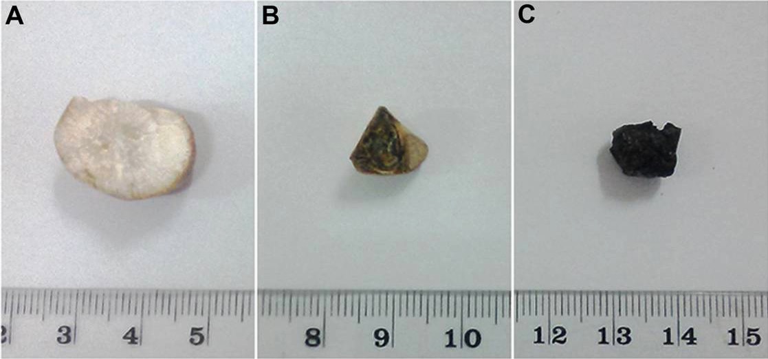
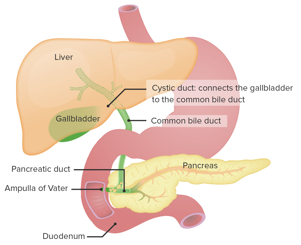
00:00 Welcome. 00:01 Today we'll discuss disorders of the biliary system specifically about gallstone diseases. 00:08 So we'll start with a case. 00:10 A 25-year-old woman with no known medical history presents to urgent care with intermittent right upper quadrant pain. 00:17 In the past 2 weeks, she has had several episodes of pain after eating which last for a few minutes. 00:22 Today, she had an episode of severe right upper quadrant pain with nausea, which resolved after 30 minutes. 00:29 She takes no medications. 00:31 Her vitals are normal, abdominal exam is unremarkable. 00:34 Her lab studies including a CBC, lipase and hepatic panel are normal. 00:39 Ultrasound of the right upper quadrant shows gallbladder stones without ductal dilatation. 00:45 So what is the best next step in management? So let's point out that she has intermittent right upper quadrant pain, after eating which typically last for a few minutes. 00:56 Her normal vitals exam and labs are all quite reassuring. 01:02 And her right upper quadrant ultrasound shows the presence of gallstones. 01:08 So, let's now discuss gallstone disease. 01:11 So there are two types of gallstone disease: First you may have "cholelithiasis" which is when you have a stone in the gallbladder and the term "choledocholithiasis" refers to a stone in the common bile duct. 01:26 So now let's review the different terminology for gallstone diseases. 01:30 I always found this to be quite confusing so we're gonna break it down step by step. 01:35 The first term is when you have a gallstone in the gallbladder without any obstruction. 01:41 This is called asymptomatic cholelithiasis. 01:45 Patients often have a normal hepatic panel. 01:48 You can detect the diagnosis by ultrasound, and treatment is just with observation. 01:55 Next if that stone in the gallbladder causes obstruction every now and then, this can lead to the development of biliary colic, or just pain that occurs intermittently. 02:06 This is what's called symptomatic cholelithiasis. 02:10 Again, they will have a normal hepatic panel. 02:12 You can diagnose them by ultrasound. 02:15 And the treatment in this case is with an elective cholecystectomy. 02:20 Lastly, you may have a stone that is now blocking the common bile duct. 02:25 So now this is choledocholithiasis. 02:29 These patients may have an elevation or cholestatic pattern elevation in the hepatic panel. 02:34 The imaging can be done with either an MRCP which we'll talk about later, or ultrasound. 02:40 And treatment is with removal of that stone with a procedure called an ERCP and a sphincterotomy. 02:50 So, how do patients with cholelithiasis typically present? We talked about the term biliary colic which is this acute onset of upper abdominal pain, usually lasting for several minutes to several hours, is often accompanied by nausea and vomiting and these episodes tend to be self-limited and occur in relation to eating. 03:16 So again, recall that this is from a gallstone becoming impacted or obstructing the gallbladder temporarily. 03:21 So patients who have cholelithiasis may either be asymptomatic or they may develop symptoms that presents with biliary colic. 03:30 The diagnosis is made by a right upper quadrant ultrasound to detect gallstones. 03:34 And treatment depends on whether or not they have symptoms. 03:37 So if they are asymptomatic, you simply observe them. 03:41 However if they have symptoms, then you perform an elective cholecystectomy. 03:45 This is just an elective surgery where we remove the gallbladder. 03:52 So there are several different types of gallstones that you may be tested on. 03:56 The first type is cholesterol or mixed gallstones. 03:59 These tend to be associated with female gender, advanced age and certain indigenous populations in the Americas. 04:06 These are shown by panel A You may also have pigmented gallstones associated with hemolysis, cirrhosis or disease of the ileum. 04:16 These are shown in panels B and C So now let's return to our case. 04:22 A 25-year-old woman with intermittent right upper quadrant pain after eating. 04:27 She has a normal vitals exam and labs and her ultrasound shows gallstones. 04:31 So, we now know that her intermittent pain associated with eating is considered biliary colic. 04:38 Her normal exam and labs are reassuring against acute cholecystitis. 04:42 And again, her ultrasound shows evidence of gallstones but no evidence of cholecystitis So, she now has fallen into the category of symptomatic cholecystitis and the best next step in management would be to refer her for an elective cholecystectomy.
About the Lecture
The lecture Cholelithiasis vs. Choledocholithiasis with Case by Kelley Chuang, MD is from the course Disorders of the Hepatobiliary Tract.
Included Quiz Questions
Which of the following is the treatment of choice for symptomatic cholelithiasis?
- Elective cholecystectomy
- Observation
- Sphincterotomy
- ERCP
- MRCP
Which of the following is the diagnostic test of choice for cholelithiasis?
- RUQ ultrasonography
- Abdominal CT
- RUQ X-ray
- ERCP
- RUQ MRI
Author of lecture Cholelithiasis vs. Choledocholithiasis with Case

Kelley Chuang, MD
Customer reviews
5,0 of 5 stars
| 5 Stars |
|
1 |
| 4 Stars |
|
0 |
| 3 Stars |
|
0 |
| 2 Stars |
|
0 |
| 1 Star |
|
0 |
La información es clara y concisa, por lo que considero que es buen material de repaso.
