Playlist
Show Playlist
Hide Playlist
Cavernous Sinuses

-
Slides 20 DuralVenousSinuses BrainAndNervousSystem.pdf
-
Reference List Anatomy.pdf
-
Download Lecture Overview
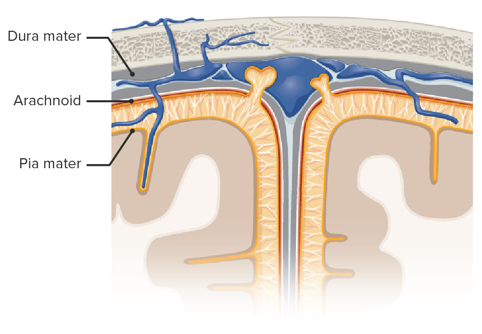
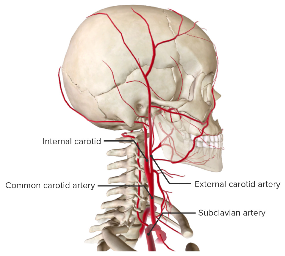
00:00 I now want to have you revisit the cavernous sinuses. The purpose of this is to understand the structures that relate to the cavernous sinuses. Then we’ll have an opportunity here shortly to emphasize the clinical relevance of these related structures. So here we’re looking at the cavernous sinuses. They are paired. The first structure related to the cavernous sinus is the internal carotid artery. These would be paired structures. So, we see it on this side of the image and on the right side of the image here as well. Traveling with the internal carotid arteries would be the carotid plexus of sympathetic nerve fibers. These are not shown in this particular image. Another structure that relates to the cavernous sinus is one of the cranial nerves. This happens to be the abducent nerve or cranial nerve number VI. 01:11 We see that slender nerve right in through here. You can appreciate the anatomic intimacy of the abducent nerve with the internal carotid artery. Oculomotor nerve, cranial nerve number III relates to the cavernous sinus. 01:27 Here, we can see that it’s located within the wall of the cavernous sinus, similarly here on the opposite side as well as you would expect. The trochlear nerve has a relationship as well to the cavernous sinus. 01:44 We can see the trochlear nerve within the wall here. The ophthalmic division of the trigeminal nerve or the ophthalmic nerve itself, V/1 is located again within the wall of your cavernous sinus. There’s one final cranial nerve that has a relationship here. It’s more inferiorly and laterally displaced. That is going to be cranial nerve V/2, which is the maxillary nerve or the maxillary division of your trigeminal nerve. Now, for some clinical correlations or considerations, first is that we’re going to explore. I want you to think about the external aspect of the face and some of the communications that can occur here with the cranial cavity. So, we’re going to explore here briefly thrombophlebitis of the facial veins. The facial veins course across the face up into the medial corner of the eye, right along in through here on either side. They’re not shown but along the green triangle here is the relationship of both of your facial veins. This green triangular area represents the danger triangle or the danger zone as it relates to thrombophlebitis of the facial veins. What are some of the considerations of thrombophlebitis of facial veins? The consideration here is that an infection because this is an inflammatory state, the infection can spread from the danger triangle internally. So if you then end up with an infection because of the communications that exist between the facial veins and the dural venous sinuses, if that infection spreads into the cavernous sinuses on either side here, you can then start to involve some of the structures that are related to the cavernous sinuses. A structure that’s particularly involved in this clinical state is the abducent nerve. If it’s affected or involved by this inflammatory state, it will cause an internal strabismus. 04:21 Other cranial nerves because of their relationships may also be involved resulting in various types of cranial nerve palsies. The last consideration is that if septic thrombosis results, this will then usually cause acute meningitis. Again, with the cavernous sinuses, we now want to consider a pituitary gland tumor and what impact this tumor might have in structures that relate to the cavernous sinuses. So here, we see the pituitary gland right in through here sitting in the sella turcica with the right and left cavernous sinuses then on the lateral aspects. Now, imagine a pituitary gland tumor. It’s expanding. It’s growing. 05:12 It grows more so laterally than it does superiorly and inferiorly because of the constrictions of superior and inferior structures. So the spread proceeds with less resistance to the lateral aspects. So what do we have here if this tumor is growing, expanding out laterally. A consideration here is the presence of your internal carotid arteries. If this tumor puts enough pressure on your internal carotid arteries, it can start to narrow them, cause those arteries to become stenotic. If there’s enough stenosis, the driving pressure to the territories that are supplied by these arteries results in an infarction. If the tumor involves cranial nerves, perhaps the abducens nerve, perhaps oculomotor, then you’ll have various forms of cranial nerve palsies as well as a result of this mass effect of a tumor.
About the Lecture
The lecture Cavernous Sinuses by Craig Canby, PhD is from the course Dural Venous Sinuses. It contains the following chapters:
- Cavernous Sinuses
- Clinical Correlations
Included Quiz Questions
Which of the following structures does NOT pass through the cavernous sinus?
- Mandibular division of the trigeminal nerve
- Maxillary division of the trigeminal nerve
- Abducens nerve
- Oculomotor nerve
- Ophthalmic division of the trigeminal nerve
Which of the following could NOT be caused by a right-sided cavernous sinus infection?
- Loss of taste to the right side of the tongue
- Right-sided ophthalmoplegia
- A fixed, dilated pupil
- Maxillary sensory loss
- Ptosis
Which of the following could be caused by a septic thrombus in the cavernous sinus?
- Acute meningitis
- Pharyngitis
- Gingivitis
- Endophthalmitis
- Vestibular neuritis
Which of the following structures in the cavernous sinus could be compressed by a pituitary tumor growing laterally?
- Internal carotid artery
- Basilar artery
- Bridging veins
- Middle meningeal artery
- Cranial nerve VII
Author of lecture Cavernous Sinuses

Craig Canby, PhD
Customer reviews
5,0 of 5 stars
| 5 Stars |
|
1 |
| 4 Stars |
|
0 |
| 3 Stars |
|
0 |
| 2 Stars |
|
0 |
| 1 Star |
|
0 |
feeling amazing after knowing everything about cavernous sinus . Its simple and quick
