Playlist
Show Playlist
Hide Playlist
Case: 6-year-old Girl Hits Her Head

-
Slides Head Trauma Epidural Hemorrhage.pdf
-
Download Lecture Overview
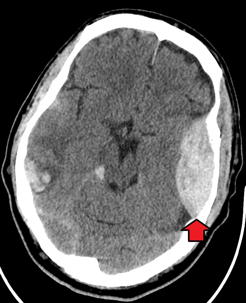
00:00 In this talk, let's discuss epidural hemorrhage. 00:05 And let's start with a case. 00:07 This is a young 6 year-old girl in her normal state of health, who was sledding in the snow with friends when she was struck from standing by a sled and falls onto the ground hitting her head. 00:19 After being a bit confused on the ground, she stands up and appears normal. 00:23 She's taken home to rest and watched closely by her parents. 00:28 Several hours later, the patient becomes increasingly sleepy. 00:32 She's taken to the emergency department, where she somnolent, difficult to arouse, has right hemibody weakness that withdraws only with pain and a CT of the head is performed. 00:43 So when you think about this case, there are a number of key features here. 00:46 First is the traumatic event, which is the nidus for this presentation. 00:51 And then there's this appearance of a lucid interval, an initial period of confusion, followed by normalization and then progressive neurologic decline. 01:00 And in terms of localization, she has a focal neurologic presentation with right hemibody weakness suggesting a left cortical left hemispheric problem. 01:11 So let's look at the imaging. 01:12 A noncontrast head CT was performed and demonstrated this lesion at the back and base of the brain behind the cerebellum. 01:20 There is this lens-shape, hyper density, extra axially outside of the brain. 01:28 So what's the most likely diagnosis? Is this an epidural hematoma, subdural hematoma, concussion or mild TBI or an intraparenchymal hemorrhage? Well, this isn't the imaging appearance of a subdural hematoma, subdurals are crescent-shaped lesions that extend beyond suture lines of the brain. 01:47 It's more common in older individuals, we can see it in young individuals, and it typically progress presents with a steady and gradual progression of symptoms. 01:58 This isn't the appearance of an intraparenchymal hemorrhage, the blood here is outside of the brain and not within the parenchyma proper. 02:05 And so this is inconsistent with IPH at least what we've seen thus far. 02:10 This is not the presentation of a mild TBI. 02:13 The patient has significant neurologic deficits intracranial hemorrhage, and so this is inconsistent with mild TBI or concussion. 02:22 And this is a typical presentation of a patient with an epidural hematoma both the clinical presentation and the imaging findings.
About the Lecture
The lecture Case: 6-year-old Girl Hits Her Head by Roy Strowd, MD is from the course Head Trauma.
Included Quiz Questions
What condition is most likely present in a patient with a traumatic brain injury who demonstrates a "lucid interval" followed by focal neurologic deficits and progressive cognitive decline?
- Epidural hematoma
- Intraparenchymal hemorrhage
- Concussion
- Subdural hematoma
- Subarachnoid hemorrhage
If imaging findings after a traumatic brain injury demonstrate hemorrhage within the brain itself, what diagnosis is most likely?
- Intraparenchymal hemorrhage
- Subdural hematoma
- Lacunar infarct
- Epidural hematoma
- Subarachnoid hemorrhage
Author of lecture Case: 6-year-old Girl Hits Her Head

Roy Strowd, MD
Customer reviews
5,0 of 5 stars
| 5 Stars |
|
5 |
| 4 Stars |
|
0 |
| 3 Stars |
|
0 |
| 2 Stars |
|
0 |
| 1 Star |
|
0 |
