Playlist
Show Playlist
Hide Playlist
Cardiac Case: 86-year-old Woman with Shortness of Breath during Annual Checkup

-
Cardiac Case 86-year-old Woman with Shortness of Breath during Annual Checkup.pdf
-
Reference List Cardiology.pdf
-
Download Lecture Overview
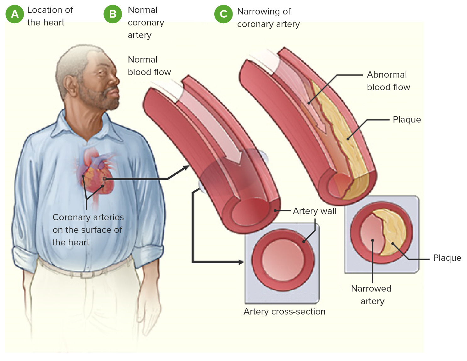
00:01 An 86-year-old woman reports during her annual checkup that she’s been feeling increasing shortness of breath when doing daily household chores and several times, she’s felt like she was going to faint during recent weeks. 00:13 She denies any chest discomfort with exertion, she’s otherwise well lives alone independently and functions well with excellent cognitive function. 00:25 She's had long standing osteoarthritis which causes her some joint pain but otherwise she’s been healthy. 00:31 On physical exam, the blood pressure is normal 140/72, heart rate normal at 84 and her peripheral oxygen saturation is also normal and the jugular venous pulse is not elevated so it suggest she’s not in heart failure. 00:45 Her respiratory sounds are clear so it suggest that there’s no fluid in her lungs. 00:50 Her heart exam is abnormal, there’s a very loud grade 3/6 ejection systolic murmur best heard at the right upper sternal border and there is no third heart sound. 01:03 Let me imitate that for you. Here’s the normal, lub-dub, lub-dub, lub-dub. 01:09 Here's what we hear in this lady’s exam, hurm, hurm, hurm, hurm. 01:15 So this is very highly suggestive of severe aortic stenosis. 01:21 All of her laboratory blood tests are normal which is good, she has normal renal function. 01:26 So here are the critical factors. 01:28 The symptom that’s critical is the short of breath which suggest that there is some either heart or lung problem. 01:35 She’s had some dizzy spells not quite fainted but close to fainting. 01:39 Her blood pressure is reasonable a little bit elevated systolic hypertension but not uncommon in a lady of this age and of course she has the murmur of aortic stenosis. 01:53 We do an electrocardiogram and it shows left ventricular hypertrophy with STT changes. 02:00 You can see here an example of a normal EKG compared to lead V6, you can see how abnormal this complex is, it’s very large R wave and a down slopping ST segment with an inverted T so called left ventricular hypertrophy with STT changes means quite significant left ventricular hypertrophy. 02:24 And here’s a slice from her echo. 02:26 The left ventricle looks pretty good, the left atrium is a bit dilated and the aortic valve is quite stenotic. 02:34 She undergoes a cauterization and has a gradient of 60 mmHg across the calcified aortic valve, this confirms the diagnosis of aortic stenosis. 02:43 Her coronary artery is fortunately only have minor atherosclerotic narrowing so nothing needs to be done there and the diagnosis, aortic stenosis quite severe due to a calcified aortic valve and she undergoes transcutaneous aortic valve replacement - that is a catheter, replacement of the aortic valve. 03:02 She’s discharged to a rehabilitation center for a week or two before returning home and we anticipate that she will do quite well.
About the Lecture
The lecture Cardiac Case: 86-year-old Woman with Shortness of Breath during Annual Checkup by Joseph Alpert, MD is from the course Cardiovascular Cases.
Author of lecture Cardiac Case: 86-year-old Woman with Shortness of Breath during Annual Checkup

Joseph Alpert, MD
Customer reviews
5,0 of 5 stars
| 5 Stars |
|
5 |
| 4 Stars |
|
0 |
| 3 Stars |
|
0 |
| 2 Stars |
|
0 |
| 1 Star |
|
0 |
