Playlist
Show Playlist
Hide Playlist
Capillaries: Overview

-
Slides 02 Human Organ Systems Meyer.pdf
-
Reference List Histology.pdf
-
Download Lecture Overview
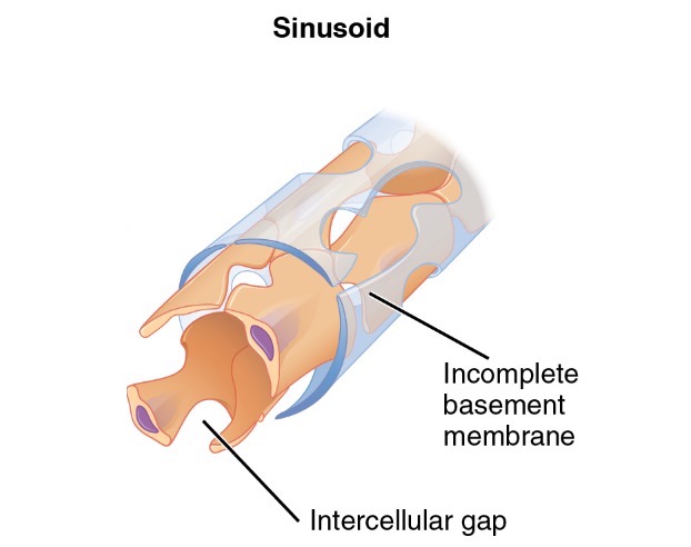
00:01 Let's have a look at our capillary bed. Capillaries are very, very hard to see when you look at sections of tissue because they're under very low pressure, if any pressure at all may collapse them. So they're almost impossible to see. Here's a diagram illustrating on the left-hand side, the nature of the capillary bed. And on the right-hand side, you can see again the image or the section that I've used previously. On the bottom left-hand side of the histological section, you can see a little small artery. It's going to get smaller and smaller to be only one or two layers of smooth muscle and become an arterial. 00:45 And on the bottom right-hand side of the image, you can see half a lumen or half a section through a venule, a small venule. Look at the thin wall. And at top, on the top of the section, you can see a structure with a large lumen, a very very thin wall. That's probably going to be a postcapillary venule or even a small venule. It's very hard to know, but just get an idea of the relative thickness of the walls of these vessels. Well, blood flows through the arterial and goes into the capillary bed. And you can see that illustrated in the diagram on the left-hand side. That arterial sends blood through branches, through very small branches, into the capillary bed where there's a lot of networks, a lot anastomosis, a lot of connections. So blood flows through that capillary bed in an enormous network or surface area, supplying all the surrounding cells and the interstitial fluid with all the nutrients that the cells need, and collecting all the waste products from the interstitium, from the cells, and then returning it back towards the heart to be dealt with by other organs. Sometimes histologists like to describe metarterioles or precapillary sphincters. 02:10 They can sometimes close off the supply of the capillary bed or blood from the arterioles, or sometimes they can close off in one area and open up in another area and divert blood directly from the arteriole side of the cardiovascular system directly back into a venule and bypass the capillary bed all together. It's called an arteriovenous anastomosis. And that occurs in some parts of the body, for instance in the dermis of skin that I've mentioned earlier. 02:50 There are sweat glands penetrating down from the epithelium, from the epidermis of the skin. Those sweat glands have a role in thermoregulation. They secrete fluid water to the surface of the epidermis where it's evaporated and that cools the body down. Well, sometimes when we overheat, when we play sport for instance, and we increase our sweating, we also like to dissipate more and more heat from the body. So sometimes the dermis of skin opens up. The blood supply to the skin is opened up by these channels opening up and sending more and more blood to the skin surface towards the skin surface. The epidermis is avascular, but there is a vast network of blood vessels within the dermis right underneath that epidermis. So in conditions, hot conditions where we want to try and dissipate body heat, we can open up these capillary channels and the heat can be dissipated across the surface. Conversely, if we're in very cold weather, we can close off that blood supply to the dermis, and therefore, not lose body heat. So these are often very important structures. But let's concentrate now on blood flow into the true capillaries. 04:17 They go to capillaries where there's this exchange, then they move into postcapillary venules and then back into the venules to be returned to the heart. 04:28 I want to concentrate now on the structure of these capillaries, which as I've emphasized is very hard to see when you look at histological sections. 04:43 So let's look at the structure of a capillary. 04:45 There are actually three types of capillaries and I'll describe them briefly here, but then I'll emphasize them more when we look at their functional role in other organ systems. 04:58 On the left hand side, you can see a picture or an image taken through connective tissue, and there are blood capillaries running through them. 05:10 Very, very thin, you can make out just the very thin walls. 05:15 You can see through them and just might get the elongated structures that represent the nuclei of the endothelial cells. 05:23 Well, probably the most common type of capillary is what we call a continuous capillary, and this is illustrated on the diagram, particularly on the bottom diagram on the right hand side. 05:38 A continuous capillary is one that I've referred to before. 05:42 This is where the endothelial cells join together to make up the capillary lining and it's joined together by very strongly occluding junctions. 05:55 These strongly designed occluding junctions include tight junctions, so the junctions between endothelial cells is very, very solid. 06:05 Very strong, nothing passes through those junctions. 06:10 And if you'll look back on your knowledge of epithelia, these occluding junctions were very important in many epithelial tissues because they restrict movement of fluid and other substances, pathogens between cells. 06:28 So, in many organs of the body, skin and particularly the brain, you have these continuous capillaries and the only way in which substances are transported across the capillary wall, is by pinocytosis, by these substances being invested by the cell, taken in by the cell, wrapped up in little membranes, and transported across the endothelial cell surface in cytoplasm. 07:04 And then it's released on the other side into the interstitium. 07:08 It's called transcytosis. 07:11 The movement of fluid and other substances by endocytosing that material, the lumen, and by those vesicles moving across and releasing the products on the other side. 07:24 That enables these capillaries to restrict the sorts of substances that they allow to pass across them. 07:33 The basal lamina wrapped around the endothelium is always continuous. 07:38 Well, another sort of capillary is one where the actual cytoplasm of these very, very thin endothelial cells have little fenestrations, little windows, little pores, and that allows substances to pass out through those pores, albeit restricted and albeit very finely controlled. 08:07 We'll see an example of these fenestrated capillaries in the kidney because they form part of the filtration component of our blood forming a glomerular filtrate. 08:21 In that situation, there's a very thin diaphragm between the fenestrations because there needs to be further control over what passes from the blood in the kidney. 08:33 But just to summarize these little capillaries, again, the basal lamina is continuous, but there are little windows or little pores within the cytoplasm, allowing substances to pass through them. 08:48 Well, the final type of a capillary, we call a discontinuous capillary. 08:57 It has a discontinuous basal lamina, sometimes we refer to this as being sinusoidal. 09:04 There are large gaps in the cytoplasm, in the lining of the epithelium of the blood vessel, the endothelium, and these large gaps are quite typical in some endocrine tissues. 09:18 They're very leaky. They allow substances to pass out of them and also into them. 09:25 And in some organs such as the spleen, cells actually pass through these large gaps. 09:32 The liver is another example of these sorts of capillaries. 09:37 And again, let me stress that we will look at these types of capillaries when we look at the different organ systems where they have a functional role.
About the Lecture
The lecture Capillaries: Overview by Geoffrey Meyer, PhD is from the course Cardiovascular Histology.
Included Quiz Questions
Exchange of fluid, nutrients and waste products occur mainly through which of the following types of vessels?
- Capillaries
- Venules
- Arteries
- Arterioles
- Veins
Which of the following is the main component of the capillary wall?
- Endothelium
- Smooth muscle
- Elastin
- Tunica adventitia
- Pericyte
Author of lecture Capillaries: Overview

Geoffrey Meyer, PhD
Customer reviews
4,0 of 5 stars
| 5 Stars |
|
3 |
| 4 Stars |
|
0 |
| 3 Stars |
|
0 |
| 2 Stars |
|
0 |
| 1 Star |
|
1 |
Excellent explanations and clear, thank you! Pretty cool you got the histology guide guy to make a lecture.
Very well done, lecturer uses excellent visual aids and makes clear explanations
Very clear explanation, understandable, good examples, clear slides and pictures.
cheap lecture slides put key points and words instead of just whole bunch of diagrams. at least label the diagrams
