Playlist
Show Playlist
Hide Playlist
Traumatic Brain Injury: Diagnosis

-
Slides Head Trauma Traumatic Brain Injury.pdf
-
Download Lecture Overview
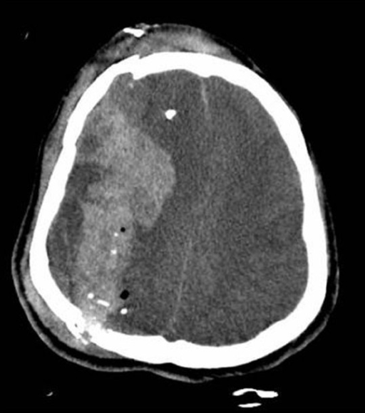
00:01 What's the mechanism of a traumatic brain injury where we can see closed head injuries where the head has been struck by something, the head strikes something that is fixed. 00:12 This can result in an acceleration-decelerate deceleration injury or injury as a result of rotational mechanisms. 00:19 And each of these mechanisms can contribute to a traumatic brain injury. 00:24 We can also see traumatic brain injuries from penetrating trauma or blast trauma. 00:30 One of the important things to think about is how the brain is injured. 00:34 And an acceleration-deceleration mechanisms, we can see two injuries to the brain. 00:40 Both a coup injury that occurs at the site of the direct impact of the brain on surrounding structures, including the skull. 00:49 And a secondary contrecoup injury which occurs at the area of the brain opposite the site of impact. 00:57 And this contrecoup injury can actually often result in more severe injuries to the brain. 01:02 And so we may see frontal and occipital injuries, we may see bitemporal and parietal injuries as a result of this coup and contrecoup injury. 01:14 We grade and scale traumatic brain injuries based on the severity of the patient's symptoms. 01:20 And we can categorize TBI as mild, moderate or severe. 01:24 And there are a number of things that I want you to think about as you're evaluating whether a patient may have suffered a mild, moderate or severe traumatic brain injury. 01:32 First we use the Glasgow Coma Scale. 01:35 Mild TBI is defined as a GCS of 13 to 15. 01:39 Moderate TBI is 9 to 12 and severe TBI as less than 8. 01:45 We also think about loss of consciousness. 01:47 With mild TBI, there is often a brief period of loss of consciousness or none at all. 01:52 The patient may maintain consciousness throughout the entirety of the traumatic event. 01:58 With moderate TBI there is frequently loss of consciousness in the range of 30 minutes with impaired a consciousness, sometimes extending out to several days. 02:09 And then with more severe TBI, patients can remain in a coma for a prolonged period of time requiring hospitalization during that. 02:17 Post-traumatic amnesia is a differentiating factor. 02:21 Amnesia after the traumatic event is typically very short or not at all. 02:24 With a mild TBI, it can last 1 to 7 days. 02:28 With moderate TBI, it may last for a prolonged period of time with severe TBI. 02:34 And we can often see some other key factors that help differentiate these three categories. 02:39 Non-severe mechanisms may be in place for mild TBI, such as a fall or an injury during practice in a sporting event. 02:47 With moderate TBI, patients may have headache, severe vomiting that may be refractory to medication interventions or alterations in their mental status at the time of the injury. 02:57 And then severe long term and even persistent neurologic deficits can result from severe TBI. 03:04 What about diagnosis? How do we diagnose a TBI or traumatic brain injury? Well, first the Glasgow Coma Scale is critical. 03:12 We often perform a pupillary examination looking for unequal pupils or anisocoria. 03:18 Anisocoria is a concerning finding and should prompt immediate workup for cause of of that abnormal pupil exam. 03:26 In particular, we think about herniation, either transtentorial herniation, uncal herniation or brainstem herniation, which could be compressing the third cranial nerves or other important structures. 03:39 In addition, in patients with neck injury, your injury to the carotid artery can result in a Horner syndrome which may lead to abnormal pupil exam, ptosis, meiosis and anhidrosis which requires imaging evaluation for possible dissection. 03:58 In addition to the physical exam, imaging is critical in patients presenting with TBI particularly moderate to severe TBI. 04:05 A CT of the head without contrast is the first line modality of choice. 04:09 We're looking for skull fractures, intracranial hemorrhage or cerebral edema. 04:12 And here we see a noncontrast head CT demonstrating an open skull fracture with underlying cerebral edema. 04:19 MRI has better diagnostic sensitivity for underlying brain pathology. 04:24 And here we see an associated MRI demonstrating a significant area of broad temporal contusion.
About the Lecture
The lecture Traumatic Brain Injury: Diagnosis by Roy Strowd, MD is from the course Head Trauma.
Included Quiz Questions
What category of brain injury is blunt trauma with a baseball bat?
- Closed head injury
- Penetrating injury
- Rotational injury
- Ischemic injury
- Blast trauma
In a patient with blunt force trauma to the frontal region of the brain, where might a clinician expect to localize the contrecoup injury?
- Occipital area
- Parietal area
- Temporal area
- Prefrontal area
- Motor cortex
What level of TBI severity is associated with post-traumatic amnesia lasting more than seven days?
- Severe
- Moderate
- Mild
- Progressive
- Regressive
What level of TBI severity is a Glasgow Coma Scale (GCS) score of 10?
- Moderate
- Mild
- Severe
- Comatose
- Normal
Author of lecture Traumatic Brain Injury: Diagnosis

Roy Strowd, MD
Customer reviews
5,0 of 5 stars
| 5 Stars |
|
5 |
| 4 Stars |
|
0 |
| 3 Stars |
|
0 |
| 2 Stars |
|
0 |
| 1 Star |
|
0 |
