Playlist
Show Playlist
Hide Playlist
Bone Formation

-
Slides 09 Types of Tissues Meyer.pdf
-
Reference List Histology.pdf
-
Download Lecture Overview
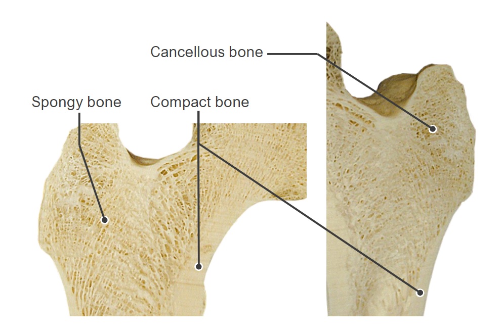
00:00 Well, that background information on bone, bone structure and the sorts of cells that are in bone and help form bone. 00:09 Let us now have a look at bone formation. It begins about the eighth week of fetal life. 00:19 It goes through into the 12th week of fetal life and then a second stage develops around the end of the second trimester. On this section, you can see the two sorts of ways in which bone forms. On the left, there is an image of endochondral ossification. On the right, is an example that was seen before in the previous slide, of intramembranous ossification where the progenitor cells are derived from mesenchyme and they turn into osteoblasts and lay down bone. So they are the two major processes. I am not going to refer to intramembranous ossification in a great deal now for the rest of this lecture because it is involved with formation of bones of the cranium and some of the jaw bones and they simply are derived from this membranous origin. What I am going to concentrate on, is the formation of bone that relates to the long bones or skeletal bones that help us to move our limbs by the action of skeletal muscles on them. So just summarize yourself what do you think are the major components of intramembranous ossification. It is important you understand that process of ossification. Well now look at the other processes. What happens in the fetals, first of all is that mesenchyme cells will develop into chondroblast, cells that lay down cartilage and will secrete type II collagen and also the matrix similar to the matrix that are described a moment ago and also the matrix I described in another lecture in this course on cartilage. And you can see in this image all the little chondrocytes surrounded by matrix. They were chondroblasts that originally derived from the mesenchyme lie down the matrix and now they're surrounded by the matrix. And they are sitting in little spaces called lacuna. You can just see the little dots that represents probably the nuclei of these chondrocytes. But during processing, when there is shrinkage of these chondrocytes these little space you see around in the hilar that we call the lacunar space is really artifact. 03:00 Those spaces are occupied by the entire cell nucleus and cytoplasm of the chondrocyte. And they can get their nutrition because their matrix allows nutrients to fuse throughout unlike in bone. Well, one of those stages the very first stage of forming a bone is that where the bone is going to be in the body. There is a cartilage model first created and that cartilage model as you see in this slide actually starts to form the rough shape of what the bone is going to be like, not the size of the eventual bone, but it's the shape. And what happens then is a whole series of processes where that cartilage template or model is then replaced by bone through a process I am now going to describe. The cartilage will grow by either interstitial growth meaning the cartilage cells within divide and they spread apart or the cartilage will grow to its proposed shape by appositional growth, which is growth of cartilage on the surface. They are the two ways in which cartilage grow. Well what happens at this stage when at about the second trimester of fetal life, that cartilage model suddenly undergoes some rather drastic changes. For a start, where the diaphysis is going to be. All of a sudden, the perichondrium that is the capsule around the cartilage template, suddenly decides it does not want to be a perichondrium anymore because it is going to turn into a periosteum and start laying down bone. It starts to get a vascular supply so that perichondrium at the future diaphysis changes to be a periosteum and that lays down bone. And you see evidence of that in this section, the very dark purply red material you see there is bone. And it forms a collar and that collar is going to outline the future diaphysis of the bone. The diaphysis of the bone that I described earlier in the lecture, we looked at the mature bone in the femur. And you can see other changes there. 05:35 You can see a cavity is formed. And within that cavity, there is a lot of cells that represent bone marrow cells. But that is the first stage, a collar and the beginnings of the diaphysis. 05:53 What happens also is that at a certain region in that cartilage template, the cartilage cells swell up. They accumulate fluid and balloon up and then they cannot get nutrition. 06:08 So the little areas of matrix between them become calcified. And as that happens, think about at the same time that collar of bone forming in the diaphysis and blood vessels coming in to that periosteum. Those blood vessels bring in osteoprogenitor cells and bone marrow cells. And those osteoprogenitor cells start to form bone and that bone is resolved by osteoclast. And that creates a cavity down the center of the developing bone, down the middle of the diaphysis. It is going to be the eventual cavity of the medullary cavity that we described earlier. So you have got a process now. We have got a bony collar and a zone of hypertrophied cartilage cells that have died and created of bone laid down upon them and then it is being resolved creating that medullary cavity as I have already described. 07:19 And that is how bone forms originally. And just to summarize this, the area you can see very dark spicules of bone being laid down by lots of osteoblasts there and they are finally resolved away. Endochondral ossification then proceeds further. What I have described results in what we have called a primary ossification center on the bottom left-hand side of destiny? What happens if this cartilage model that is being invaded by osteoprogenitor cells and has had the center taken away by the bone being developed, and then resolved. 08:15 That hypertrophied area becomes the primary ossification center. And then from the second trimester of fetal life, right and through adulthood, that primary ossification center is going to contribute to the lengthening of the bone until finally at the end of puberty that will seize and the bone length is then determined.
About the Lecture
The lecture Bone Formation by Geoffrey Meyer, PhD is from the course Bone Tissue.
Included Quiz Questions
What type of collagen plays a major role in endochondral ossification?
- Type II
- Type I
- Type III
- Type IV
- Type V
Endobronchial ossification leads to the formation of what?
- Primary ossification center
- Secondary ossification center
- Osteoid seam
- Epiphyseal cartilage
- Periosteal collar
At approximately what fetal age does bone development initiate at primary ossification sites?
- 8ᵗʰ week
- 4ᵗʰ week
- 2nd week
- 1st week
- During the third trimester
Cartilage matrix is laid down by which of the following types of cells?
- Chondroblasts
- Osteoclasts
- Fibroblasts
- Osteoblasts
- Osteocytes
Author of lecture Bone Formation

Geoffrey Meyer, PhD
Customer reviews
3,0 of 5 stars
| 5 Stars |
|
1 |
| 4 Stars |
|
0 |
| 3 Stars |
|
0 |
| 2 Stars |
|
0 |
| 1 Star |
|
1 |
Greatly assisted me in identifying the structure and function of bone organelles
So far this lecture series has been good but this particular lecture is so hard to follow. The explanations aren't clear, and the professor not pointing out anything is a problem. This has been pointed out in previous lectures as well, and the explanation given is that it's better for us if we try to find it ourselves. This is the first time we are coming across structures like this. It would be better if the professor at least points out an example to us for the first time and then allows us to identify it ourselves later. Otherwise, these lectures have been good except for this one.
