Playlist
Show Playlist
Hide Playlist
Bone Cells
-
Slides 09 Types of Tissues Meyer.pdf
-
Reference List Histology.pdf
-
Download Lecture Overview
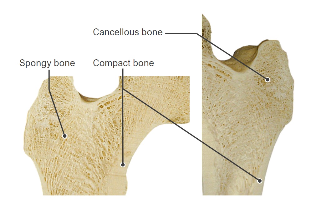
00:01 I want to now just summarize bone cells. If we look at again fetal tissue here, you see some mesenchyme and you will also see some little blastemas or little areas, where bone has started to form. The bone is that purply component you see there and within that purply component in the matrix, you can see little embedded osteocytes. 00:30 They have been lying down the bone as osteoblasts and when they get surrounded by matrix they then osteocytes. 00:39 And during their development, they all touch hands with each other. They maintain contact with each other in fine little canaliculi, and that enables nutrition to pass through these little canaliculi to all the osteocytes embedded in matrix. Endosteal cells and periosteal cells are very important components of bone. Endosteal cells are going to line all the small cavities of the bone including all the surface area of that spongy bone I showed at the very start of this lecture. The periosteal cell is going to be on the outside of the bone, the capsule around it. And they are osteogenic. If bone is damaged, those cells can then revert to be osteoblasts and repair the broken bone. Here a mesenchyme cell has developed or differentiate into an osteoprogenitor cell, and it will move to the surface of the bone you can see here and develop into osteoblasts and begin to lay down bone. And we will get back to that in a moment. The osteoblasts in this picture, are those dark stained nuclei cells sitting on the bright clear pink surface of this developing piece of bone. 02:10 Those osteoblasts have developed from those osteoprogenitor cells I described earlier and they have moved to the surface and they are lying down bone. They have been told to just develop bone from this mesenchymal type origin. You can see the dark pink calcified matrix of the bone. It is still immature bone, but you can see a very pink little line underneath the osteoblasts. That is called osteoid. It is bone that is yet to be mineralized. 02:46 So it has a different staining characteristic. But those osteoblasts will gradually mineralize that bone and the osteoblasts will be surrounded by that bone become osteocytes like the one you see here already. Those osteoblasts are secreting collagens type I plus matrix proteins and also vesicles that help with the mineralization process. Here is a lovely picture of an Australian bottlebrush flower. I put it in this lecture to illustrate the structure of a very important component of the matrix and that is the glycoaminoglycan aggregates. They are just like the red components of this bottlebrush flower you see, coming out from a central stem. Just like you see when you look at the molecular structure of these aggregates, these proteoglycan aggregates in the matrix. And those fine little red structures projecting from the central stem with little tiny yellow dots on them, they are the glycoaminoglycans. And they are negatively charged so they can accomodate. They spread out from each other. They repel each other and therefore create large spaces for water. And then allows the matrix, particularly of cartilage, to be very gel like and be very firm and rigid even though it is not calcified or even mineralized. These proteins do a similar thing in bone, they help to embed different components of the matrix and then that matrix becomes highly mineralized. One of the things that happen also is that all these matrix proteins as many of them that maintain a certain array or design of the matrix. You see a picture here of a component of a matrix of a bed, all those steel structures, all those little linked structures represent the way in which certain proteins link all the collagen type I together to form a very strong structure in the bone. And then all the material within that is going to be other matrix components that becomes very mineralized. It gives bone its strength. Here is a section through the osteon or a number of osteons. It is a ground section of bone. You can see osteocytes stained little black structures there embedded in their lacunar space and you can see some very fine black lines. They are the canaliculi, housing processes from these osteocytes. You know bone as well as cartilage, these osteocytes are also involved in mechanotransduction processes. They can feel like on the springs of the mattress I showed you earlier, they can feel different pressures. You know yourself when you are lying on a soft mattress or a hard mattress. It gives you some indication of what the composition of the matrix is. Bone cells do the same thing. They can monitor firmness of the matrix, softness of the matrix and the force that's imposed on that matrix and therefore accordingly adjusts the matrix to be able to withstand those forces and maintain the matrix in a proper chemical and fibrous constitution to actually support the role of being supportive. And just to show you some of the endosteal cells of the very central of the Haversian canal and then you see those look canaliculi that provides nutrients from the endosteal cells in the Haversian canal to all the osteocytes embedded in the matrix. 06:55 Finally, one important cell is the osteoclast shown in this slide. So at the top of this bony spicules that are being formed, those are macrophages derived cells, they don't come from an osteoprogenitor cell. They are multinucleated. They are large complexes and they eat away bone and they can liberate calcium into the blood. So they are very important in maintaining calcium levels and they are acted on by a couple of hormones that I will describe towards the end of this lecture. They sit in these little spaces called Howship's lacuna and then as I said start to dissolve away all the matrix.
About the Lecture
The lecture Bone Cells by Geoffrey Meyer, PhD is from the course Bone Tissue.
Included Quiz Questions
Which of the following regarding the endosteum is MOST ACCURATE?
- It lines the medullary cavity of long bones.
- It lines the outside surface of long bones.
- It is the major cellular component of bone.
- It breaks down bone tissue.
- It lines the epiphysial plate.
Which of the following is the predominant type of collagen in bone?
- Type 1
- Type 2
- Type 3
- Type 4
- Type 5
Which of the following is a protein constituent of the bone matrix?
- All of the other answer options are constituents of the bone matrix.
- Glycoproteins
- Proteoglycans
- Collagen
Osteoclasts are derived from which of the following types of cells?
- Macrophages
- Fibroblasts
- Neutrophils
- Osteoblasts
- Osteophytes
Customer reviews
5,0 of 5 stars
| 5 Stars |
|
1 |
| 4 Stars |
|
0 |
| 3 Stars |
|
0 |
| 2 Stars |
|
0 |
| 1 Star |
|
0 |
Great lecturer, can't recommend enough. I like his enthusiasm and clarity.


