Playlist
Show Playlist
Hide Playlist
Blood: Neutrophils

-
Slides 13 Types of Tissues Meyer.pdf
-
Reference List Histology.pdf
-
Download Lecture Overview
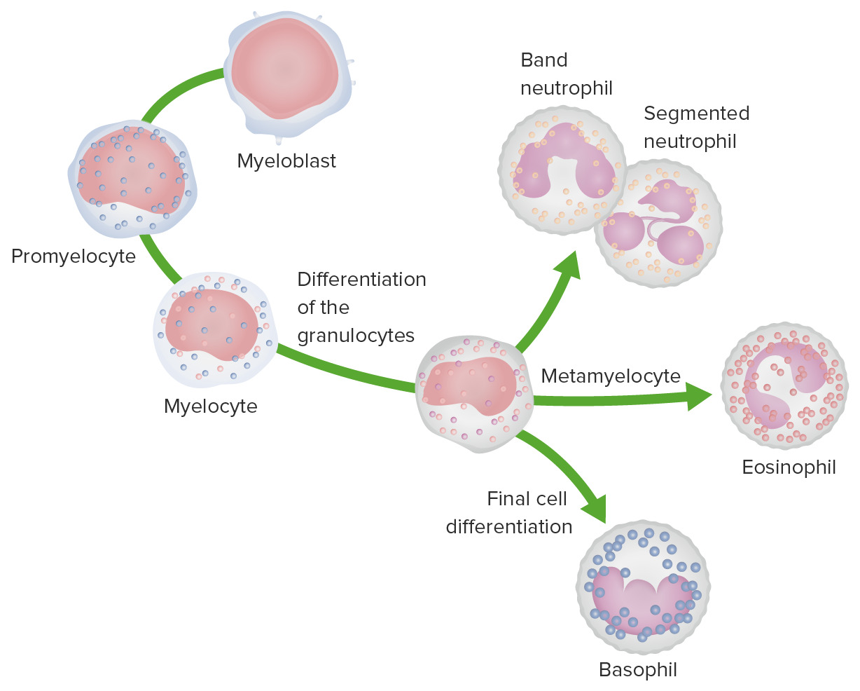
00:01 Let us look at the neutrophil first. 00:03 It is called a polymorphonuclear neutrophil or simply we often refer to them as being polymorphs. 00:12 Have a look at the cell in the middle image, right in the middle that is a neutrophil. 00:17 It has got a very strange-shaped nucleus. The nucleus has a number of lobes, dark blue stained components and each of those lobes are joined by a little tiny thread of part of the nucleus. It is polymorphous nuclei differentiate nuclear lobes and that is why we call it that names as I've mentioned. Across on the right-hand side, you can see the components of the cell under the electron microscope, you can see in those electron micrograph, a number of little granules if you look very carefully and they're reflected by specific staining that we can do in the light microscope image you see. In the case of neutrophils, it is that even though you can see these granules within the picture on the right, the electron micrograph of these cells, it does not take up stain. There are three sorts of granules that I will explain in a moment within these neutrophils, but unlike the two other granulocytes, it just does not take up the stain that we use in normal blood smears. So that is why it is called neutrophil. And it has a label. It says it lacks specific staining in the cytoplasm. 01:38 Look at the cytoplasm and it is just a very clear pink shade. Well these neutrophils are very very important because they are phagocytes. They travel around the blood, then they enter into tissue spaces where there happens to be perhaps an invading pathogen, bacteria etc. 02:00 So their job is to go in first and to mop up that debris and start to break it down. 02:06 They are the first cell to arrive at the site of inflammation and they have three sorts of granules. Some of the granules such as the specific granules are designed or at least contain antimicrobial peptides or enzymes that break down these microbes that they are attack and surround. Other granules such as the azurophilic granules, they contain lysosomes. 02:37 Lysosomes are used by the cell to breakdown the products that they've digested. 02:46 They ingest these products by digest them and they use the lysosomes to eventually break down those further components to be almost totally nothing within the cell except maybe a little bit of undigested product that they will eventually eliminate. So azurophilic granules refer to lysosome contain these hydrolytic enzymes. In the neutrophil, we also have tertiary granules. 03:15 Remember I said the neutrophil are the first cell to arrive at the site of inflammation. 03:22 And therefore they need to have a mechanism to be able to burrows through connective tissue. 03:28 So there's tertiary granules contain different sorts of enzymes such as collagenases, they allow them to move through the connective tissue very readily and therefore reach the site of infection. You can see the nucleus on the right-hand side, the electron micrograph picture, it has got a lot of heterochromatin and it is very hard to see the euchromatin when you look at a light microscope image of these cells as you see in the center. 04:04 The heavy amount of heterochromatin really indicates that these cells are not doing their job when you see them in blood. It is not until they move into the connective tissue as I have mentioned before that they actually do their job and become activated and therefore they'll become more euchromatic, which is a way of looking at the cell and identify whether it is very active or not.
About the Lecture
The lecture Blood: Neutrophils by Geoffrey Meyer, PhD is from the course Blood.
Included Quiz Questions
Which of the following cells is the most abundant type of granulocyte and the primary cell involved in chemotaxis?
- Neutrophils
- Basophils
- Lymphocytes
- Erythrocytes
- Eosinophils
Which of the following types of granules are present in neutrophils?
- All the types of granules described in the other answer options may be present in neutrophils.
- Specific
- Tertiary
- Azurophilic
Which of the following organelles is primarily involved in digesting cellular debris?
- Lysosomes
- Endoplasmic reticulum
- Ribosomes
- Golgi bodies
- Mitochondria
Author of lecture Blood: Neutrophils

Geoffrey Meyer, PhD
Customer reviews
5,0 of 5 stars
| 5 Stars |
|
5 |
| 4 Stars |
|
0 |
| 3 Stars |
|
0 |
| 2 Stars |
|
0 |
| 1 Star |
|
0 |
