Playlist
Show Playlist
Hide Playlist
Benign Liver Disease: Hepatic Steatosis, Cavernous Hemangioma and Hepatic Cyst

-
Slides Benign Liver Disease.pdf
-
Download Lecture Overview
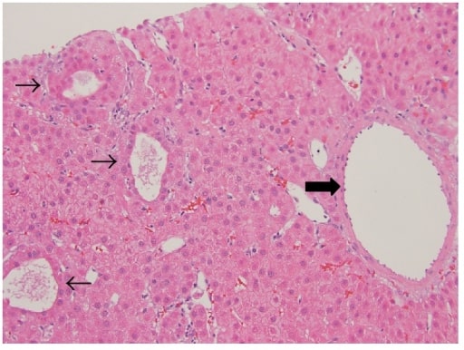
00:01 So in this lecture we will be discussing benign liver disease. 00:05 It's important to recognize benign liver disease findings so that you can help differentiate it from malignant liver disease and malignant liver lesions. 00:12 So let's take a look at these CT images. 00:16 This patient came in and had an abdominal and pelvic CT scan and we saw this lesion within the liver as an incidental finding. 00:23 Take a look at these images and see what you recognize. 00:26 As we go through the lecture keep these in mind and we'll go back to it at the end. 00:31 So benign liver abnormalities can be both focal or diffuse, focal abnormalities include cavernous hemangioma, hepatic cyst, focal nodular hyperplasia and hepatic adenoma. 00:45 Those are the most common benign liver abnormalities that are focal. 00:47 There can be multiple diffuse liver abnormalities as well, including hepatic steatosis which is also known as fatty liver, hepatitis metabolic liver disease, iron overload and autoimmune disease of the liver. 01:00 The one that will be discussing today of the diffuse category is the hepatic steatosis because that's really the only one that can be diagnosed just based on imaging. 01:09 So let's take a look at hepatic steatosis. 01:13 It's a very common diffuse benign abnormality of the liver. 01:17 It's caused by accumulation of fat in the hepatocytes and it could be diffuse or focal. It’s usually seen as a diffuse abnormality but occasionally it can be focal as well, and often is associated with either diabetes or obesity. 01:32 So this is an example of an ultrasound in a patient that has hepatic steatosis. 01:38 You can see a diffusely echogenic slightly heterogeneous liver. 01:42 This right here I'm outlining is the liver. 01:46 Usually, the liver parenchyma should be approximately equal to that of the renal cortex which is right here. 01:53 On this image you can see that the renal cortex is actually quite a bit darker than the liver and that tells you that there’s a hepatic steatosis. 02:01 Hepatic steatosis can also be diagnosed on MRI. 02:07 These are two MRI images demonstrating hepatic steatosis. 02:11 So in this first image we have the liver with a normal appearance. 02:16 A little bit brighter than the spleen, here we have the liver and here we have the spleen, on opposed phase imaging the liver demonstrates loss of signal. So again here's the liver, it's a little bit darker than it was on the in phase image over here. 02:32 And the spleen remains approximately the same. 02:36 So hepatic steatosis is best evaluated on chemical shift imaging on MRI and what we just saw the in phase and the opposed phase is an example of chemical shift imaging. 02:47 So the in phase imaging demonstrates normal signal intensity of lipid containing structures, while the out-of-phase or opposed phase imaging results in loss of signal in lipid containing tissues. 02:57 So chemical shift imaging it's really based on the very magnetic properties of lipid and water and it's very useful in identifying structures that contain lipid. 03:06 So when we discussed focal liver lesions, the most common that comes to mind is the cavernous hemangioma. 03:13 It's usually solitary, it may be multiple and if it's less than 2 cm in size is called a capillary hemangioma. 03:22 Essentially, a cavernous hemangioma and a capillary hemangioma are pathologically the same, it's just a difference in the size. 03:27 So let's take a look at what a cavernous hemangioma looks like on imaging. 03:32 Here we have multiple CT images and then an ultrasound images demonstrating the imaging characteristics of a cavernous hemangioma. 03:40 Will go into detail of each one of these in just a minute, but you can see here that we start off with a non-contrast examination. 03:47 So this is a multi-phase study. We then have an arterial phase and the portal venous phase. After that we have the ultrasound, so let’s go into a little more detail. 03:57 So on CT, a cavernous hemangioma is hypodense on the noncontrast images. 04:02 It demonstrates nodular peripheral enhancement on the early postcontrast imaging, and then it gradually becomes isodense overtime and then hyperdense on delayed imaging. 04:11 So let's take a look at the CT images here. 04:14 This is an example of our noncontrast image and you can see the lesion is actually hypodense on this noncontrast image. 04:22 It almost looks like a little cyst, this is normal liver parenchyma here but as we give contrast on the arterial phase, this is a coronal arterial phase CT scan, you can see that there is peripheral nodular enhancement. 04:36 You can see little nodules all around the periphery of this lesion. 04:39 And then on the delayed images you can also see that it's starting to fill-in, you have a little more of this nodularity around the periphery and you have a little more enhancement within the lesion as well. 04:50 On ultrasound the lesion is diffusely hyperechoic. 04:55 So let's take a look. Here we have an ultrasound image of the liver. 04:59 I'm outlining the entire liver here, and the lesion is right here diffusely hyperechoic. 05:08 This is a very non-specific finding, when you see an echogenic liver or hyperechoic lesion within the liver, you can think of cavernous hemangioma but you really do need to perform a CT or an MRI for better evaluation of this. 05:21 On MRI the lesion appears hypointense on T1. 05:25 It's hyperintense on T2. And the CT it demonstrates nodular peripheral enhancement on early postcontrast imaging. 05:33 And then gradually becomes hyperintense on delayed imaging. 05:37 So the imaging characteristics are very similar to that seen on CT. 05:40 So let's talk about hepatic cyst. This is also another very common finding that's found within the liver. 05:48 A hepatic cyst is a well circumscribed lesion and it has fluid characteristics. 05:52 So you can see here within the liver there are two hepatic cysts. 05:56 They are very well circumscribed, they’re hypodense and after contrast administration they do not enhance. 06:03 So they appear very similar initially to hemangioma. 06:06 However, after contrast administration the hemangioma will demonstrate nodular enhancement while hepatic cyst shouldn’t demonstrate any enhancement and that's how you differentiate the two.
About the Lecture
The lecture Benign Liver Disease: Hepatic Steatosis, Cavernous Hemangioma and Hepatic Cyst by Hetal Verma, MD is from the course Abdominal Radiology.
Included Quiz Questions
Which of the following is NOT a focal liver disease?
- Hepatitis
- Hepatic cysts
- Nodular hyperplasia
- Hepatic adenoma
- Hepatic abscess
Which of the following is FALSE regarding hepatic steatosis?
- There is the accumulation of fat in the intercellular space.
- On ultrasound, the liver appears diffusely echogenic and slightly heterogeneous.
- It is a common abnormality of the liver.
- It is most commonly diffuse but may also be focal.
- It is often associated with diabetes.
Which of the following statements help in differentiating a hemangioma from a hepatic cyst?
- After contrast administration, a hemangioma shows nodular enhancement while a hepatic cyst does not.
- A hemangioma appears as a well-circumscribed lesion while a hepatic cyst is more irregular.
- A hemangioma is a benign lesion of the liver whereas a hepatic cyst has malignant features.
- A hemangioma appears hypodense on a CT scan while a hepatic cyst appears hyperdense with calcifications.
- A hemangioma is filled with clear fluid while a hepatic cyst can contain solid components including teeth and hair.
Author of lecture Benign Liver Disease: Hepatic Steatosis, Cavernous Hemangioma and Hepatic Cyst

Hetal Verma, MD
Customer reviews
3,0 of 5 stars
| 5 Stars |
|
0 |
| 4 Stars |
|
0 |
| 3 Stars |
|
1 |
| 2 Stars |
|
0 |
| 1 Star |
|
0 |
organization of the MRI section is confusing. It would have been better to show the definitions and the pictures at the same time...instead of using terms not previously introduced...and then switching to the definitions, but now the viewer can't see the image which demonstrates the terms. Instead, I had to look up the terms on my own and then I could better understand the lecture. I also had to spend time going back and forth with the slides and transcript/video to better understand. In short, the organization of this section could have been done better.
