Playlist
Show Playlist
Hide Playlist
Benign Liver Disease: Hepatic Adenoma and Focal Nodular Hyperplasia

-
Slides Benign Liver Disease.pdf
-
Download Lecture Overview
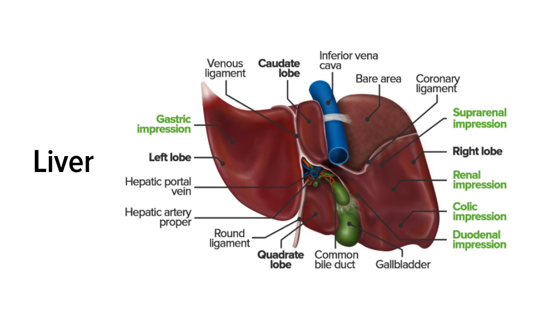
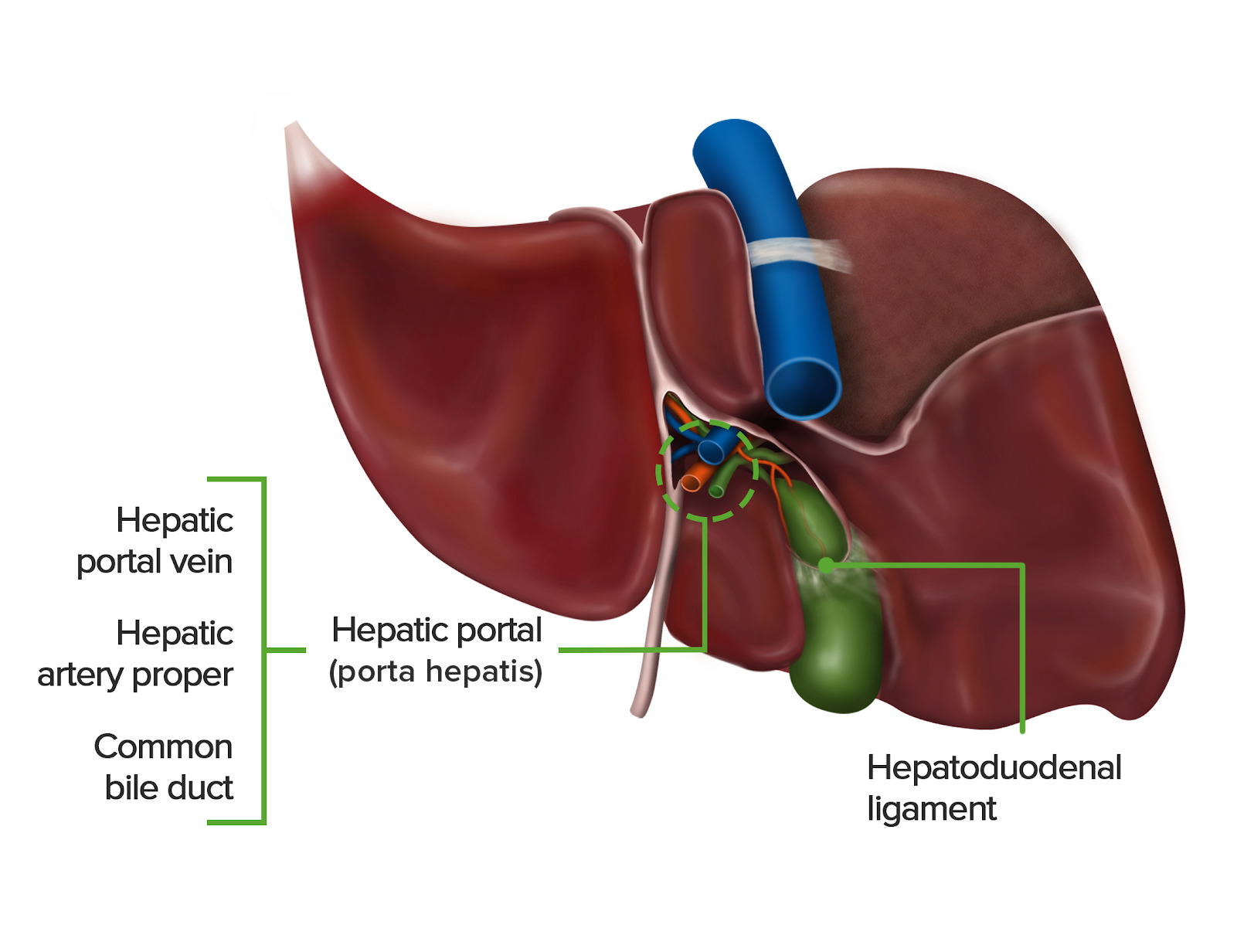
00:01 Hepatic adenoma, these are slightly less common but can be found particularly in women of childbearing age. 00:08 The most common location is subcapsular and it's found within the right lobe of the liver. 00:13 Often these can contain fat, necrosis, calcifications and hemorrhage and can appear very heterogeneous because of these findings. 00:21 The development is related to estrogen containing compound such as birth control pills. So women of childbearing age that use birth control pills are the most likely to get these lesions. 00:31 And they can actually result in severe hemorrhage which can require surgery. 00:35 All right, so let's look at some CT and MRI images that demonstrate the imaging characteristics of a hepatic adenoma. 00:42 We have arterial and portal venous phase CT scans here and we have MRI scans both demonstrating characteristic imaging of an adenoma. 00:51 Let's take a look at this in a little bit more detail. 00:54 So on a CT scan and adenoma present as a heterogeneous mass, which enhances on the early postcontrast images and then washes out early. 01:04 So this an arterial phase CT scan, you can see a very subtle finding, anteriorly within the liver and there is very subtle arterial enhancement in a portion of this finding. 01:16 The remainder of this doesn't enhance so it's somewhat heterogeneous. 01:20 On the portal venous phase the contrast washes out and now the entire lesion appears hypodense. 01:27 Again, slightly heterogeneous with this portion of it being a little more hyperdense than this portion. 01:33 On an MRI and adenoma is mildly T2 hyperintense and T1 hypointense and again it appears heterogeneous. 01:41 There's enhancement on the early postcontrast images and early washout similar to that seen on CT, and when you look at opposed phase imaging, their signal drop out because of the fat content within the lesion. 01:53 So here's an in phase and an opposed phase MRI image. 01:58 You can see the lesion right here, it's hypointense on the MRI in phase. However, in the opposed phase that actually loses even more signal and becomes more hypointense and this is again characteristic of an adenoma, because it does contain areas of lipid. 02:14 On a T2 weighted MRI image, the adenoma appear slightly hyperintense. 02:21 On an ultrasound, it's a very nonspecific heterogeneous mass. 02:25 So again, anytime a mass is seen on ultrasound, it really needs a CT or an MRI with contrast for better characterization. 02:32 Focal nodular hyperplasia is another lesion that?s somewhat uncommonly found within the liver. 02:41 It results from an anomaly of the arterial blood supply which results in hyperplastic growth of the liver parenchyma. 02:47 Oral contraceptives may promote growth, but they don't cause the development of it which is unlike an adenoma where all oral contraceptives actually do cause the development. 02:58 These can occur anywhere within the liver. 03:00 So an ultrasound, a focal nodular hyperplasia lesion appears as a homogeneous isoechoic mass that has a hypoechoic central scar. 03:09 And on CT it actually present an isodense mass, which is difficult to see on the non-contrast images. 03:16 It has intense homogeneous enhancement on the postcontrast imaging and the key feature of a focal nodular hyperplasia, is that it has a central non-enhancing scar. 03:26 Gradually this become isodense on delayed imaging with a central scar becoming more hyperdense. 03:32 On MRI, these are isointense on T1 and T2 weighted images and the central scar is dark on both T1 and bright on T2. 03:42 There's homogeneous enhancement on the postcontrast imaging, and gradually this becomes isointense on delayed imaging with a central scar becoming a little more hyperintense. 03:52 So let's take a look at an example. 03:54 Here we have a T1 contrast enhanced study and you can see that there is homogeneous enhancement of the lesion. 04:02 However, the central scar is hypointense which is characteristic. 04:07 On T2 weighted imaging, it's somewhat isointense to the rest of the liver parenchyma, but the central scar is actually slightly hyperintense. 04:16 Again, characteristic of a focal nodular hyperplasia. 04:19 So let's go back to the case that we looked at initially, we have 3 MRI images and we have the first one which is a precontrast T1 image that demonstrates a liver lesion here. 04:31 The second image is an axial postcontrast image, and the third is also a postcontrast image. 04:37 However, it's done with a 10-minute delay. 04:39 So what is your differential diagnosis here? Will take a look at each of this in a little more detail. 04:53 So in this image on the initial precontrast image, you can see that there is peripheral enhancement. 04:58 However, it's heterogeneous because the central portion is really not enhancing. 05:02 On the delayed images the entire lesion is now filled in with contrast. 05:07 So this represents a cavernous hemangioma. 05:11 It's hypointense on the T1 precontrast images. 05:14 It has peripheral nodular enhancement on the early postcontrast image and then on the delayed images there's fill-in and this is typical again of a cavernous hemangioma, which is a benign finding and no further work of needs to be performed. 05:28 So we've gone over multiple different benign liver lesions as well as diffuse liver disease. 05:33 Hopefully this will help you as we go forward with further lesions within the liver to help you characterize what's benign and what's malignant.
About the Lecture
The lecture Benign Liver Disease: Hepatic Adenoma and Focal Nodular Hyperplasia by Hetal Verma, MD is from the course Abdominal Radiology.
Included Quiz Questions
Which of the following imaging features are characteristic of hepatic adenoma?
- On CT, it appears as a heterogeneous mass with enhancement on early post-contrast images.
- On ultrasound, it appears as a homogenous solid mass.
- On MRI, there is no washout seen.
- On CT, there is no enhancement on early post-contrast images.
- On ultrasound, it is an isoechoic mass with a hypoechoic central scar.
Which statement is FALSE regarding hepatic adenoma?
- Most commonly seen in postmenopausal women
- Most common location is subcapsular in the right lobe of the liver
- There is an association with birth control pills.
- Often contains fat necrosis, calcification, and hemorrhage
- Can result in severe hemorrhage requiring surgery
Which of the following is FALSE regarding focal nodular hyperplasia?
- It most often occurs in the center of the liver.
- It results from an anomaly of the arterial blood supply.
- It is a hyperplastic growth of the liver parenchyma.
- Oral contraceptives can promote the growth of the lesion.
- It has a hypoechoic central scar seen on an ultrasound.
Author of lecture Benign Liver Disease: Hepatic Adenoma and Focal Nodular Hyperplasia

Hetal Verma, MD
Customer reviews
5,0 of 5 stars
| 5 Stars |
|
5 |
| 4 Stars |
|
0 |
| 3 Stars |
|
0 |
| 2 Stars |
|
0 |
| 1 Star |
|
0 |
