Playlist
Show Playlist
Hide Playlist
Auscultation of the Lungs: Breath Sounds

-
Reference List Physical Examination.pdf
-
Download Lecture Overview
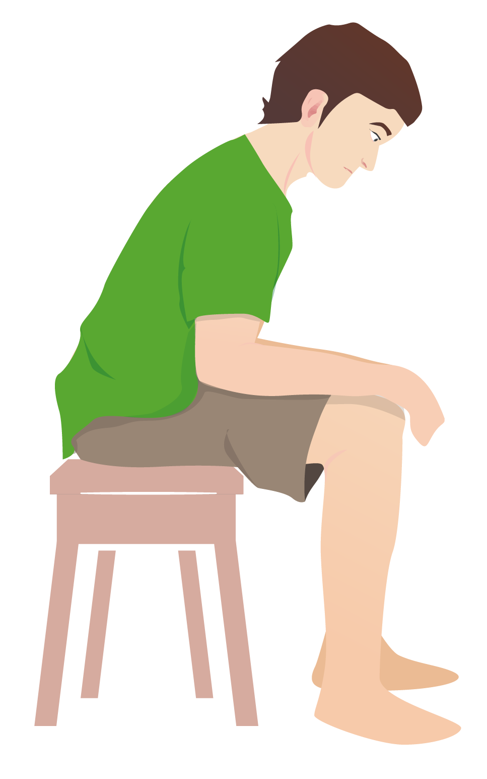
00:01 So before we put our stethoscopes on the chest of our patient, it's good to just revisualize the actually anatomy of the lobes, particularly the right lobes of the chest. 00:12 So, we have the patient lift up their elbow and put their hand on their head, you'll find the lower border of the scapula and that's good indication of approximately where the border is between these, the upper lobe of the right lung and the lower lobe of the right lung. 00:28 So I'm going to mark that here and I'm going to walk around and you can turn for me, please? And that line essentially goes all the way down to the sixth intercostals space, so that's basically the border of the -- or the upper border of the lower lobe. 00:48 And the middle lobe starts around the 5th -- the 4th intercostals space, which I know is there because the external notch marks just above where the second intercostals space is and so I can walk down and follow this line to right around here. 01:03 You can put your arm down, Shaun. 01:05 This highlights how the middle lobe of the lung can be difficult to find if you're not specifically listening in the axilla or in the anterior chest, so if you're looking for a right middle lobe pneumonia which is actually somewhat common in the setting of aspiration, you're going to miss it if you're aren't very deliberate about listening in the axilla. 01:23 With that, let's start examining our patient. 01:27 So now that we've reoriented ourselves to basic lung anatomy with the upper lobe, the middle lobe, and the inferior lobe as shown here, and keep in mind that on the left side of the chest there's just an upper lobe and a lower lobe and then a smaller lingula in the middle. 01:44 Now, we're going to talk about the different kind of things you're actually trying to find when you listen to the lungs, and the first thing I'm going to talk about is just basic breathe sounds and the types of breath sounds that you can auscultate with your stethoscope, again, the diaphragm, are either vesicular, bronchial, or something in the middle, which, you guessed it is called bronchovesicular. 02:04 So let's talk about the difference between these kinds of sounds. 02:08 I encourage you to actually take a listen to yourself or if you're with somebody else, you guys can listen together and just start by putting your stethoscope on the lower lung field in the back, and listen and describe what you hear. Take some full deep breaths for me, Shaun. 02:28 Deep breaths through your mouth. 02:32 So vesicular breath sounds are light, feathery and you may also note that inhalation is somewhat louder than exhalation. 02:43 The exhalation you may hear the beginning of it, but it trails off very quickly. 02:47 That's the sound of laminar flow through the small air ways in the peripheral lung tissue. 02:58 Now, I want you to contrast that with the sound that you might hear just lateral to the sternum and I encourage you to make sure you listen to the right side of the sternum because on the left, of course, you're going to hear the heart which will confound things. 03:10 So just listening to the right side of the sternum either on yourself or again on a friend, take a few more deep breaths. 03:25 Now, it's a subtle distinction but you'll note that there's the beginning of something that we would call bronchial breath sounds. 03:32 While you're still hearing that feathery light vesicular breath sounds in the background, you're also now starting to hear something that's a little coarser, a little bit more robust and rougher around the edges. 03:44 Still you're hearing more inhalation than exhalation but there's the beginning of a transition point there and that's because I'm listening over the main stem bronchi in the large airways of his lungs in the anterior chest. 03:57 But the next step is we're going to listen to the trachea, so put your stethoscope right over somebody's trachea in the anterior cartilage there and again, take a few deep breaths through your mouth for me, please, Shaun. 04:17 And again, you can do this on yourself or you can do it with somebody else. 04:20 And now, what we're hearing is very coarse, harsh breath sounds and you'll note the inspiration and exhalation are essentially identical in volume and pitch. 04:32 There's also less of a coarse of noise, it really is moe of a specific pitch that you're hearing and this is characteristics of tracheal breath sounds, but it's basically representing or another way of mimicking so called bronchial breath sounds. 04:47 Now vesicular breath sounds is normal, you should hear those in all the peripheral lung fields. 04:51 Bronchovesicular should only be heard here at the areas just lateral to the sternum, but bronchial breath sounds, what we were just hearing over the trachea is not normal, it's abnormal anywhere in the chest, you should not be hearing bronchial breath sounds, because what they represent is essentially the sound of air moving through the upper airways but going directly through some sort of solid tissue to the chest wall where you're listening with your stethoscope, without any of those light feathery vesicular breath sounds along the way, which tells you that there's something that is preventing air passing through the normal lung parenchyma and producing those light feathery sounds. 05:36 And that is typically going to occur with either a cancer, a large mass in the chest; or potentially a sucked in lobar pneumonia which is just transmitting those vibrations in the large airways all the way through the chest wall to your stethoscope. 05:53 So now in addition to describing the type of breath sounds you're hearing, vesicular, bronchovesicular or bronchial, you're going to want to characterize the amplitude of those breath sounds and there's a variety of different scoring systems that people use. 06:06 My approach is fairly straight forward, I simply describe the breath sounds as either full, which would be normal, moderate, fair, poor, or absent; and you can imagine that folks who have advanced emphysema with a very low FEV1, they may have a baseline only fair to poor breath sounds, you can hardly hear anything moving in the chest at all even though they appeared to be otherwise oxygenating reasonably well. 06:34 So be sure to make sure you document that full vesicular breath sounds or moderate bronchovesicular breath sounds in different areas of the chest. 06:43 In addition, you also want to look for adventitious breath sounds. 06:46 These are the sounds that are in addition to your simply description of the breath sound themselves, you're looking for things like crackles, rhonchi, and wheezing. 06:57 Crackles is a somewhat of a vague term, but it actually is describing the particular acoustics of what you're hearing, and we typically describe crackles as either fine or dry crackles, or wet also know as coarse crackles. 07:12 And fine, dry crackles we associate with interstitial pulmonary fibrosis and things of that nature that is more of a chronic indolent progression of disease and they can really be replicated by just pulling pieces of velcro apart. 07:24 They're very fine crisp, there's many different crackles that are occurring within a short span of time, and they have a higher pitch in general. 07:34 In contrast wet crackles, so called because they're often times associated with heart failure with just a little bit of extra fluid in the alveoli and they're snapping open when you take a deep breath, those wet crackles are fewer in number, they are a little bit higher in pitch, they're a little bit longer and you're going to correlate them with how high they go up in the back of the chest that may give us a sense as to how significant somebody's heart failure may be. 08:02 The next part of adventitious breath sounds is rhonchi and rhonchi is essentially are non-specific findings that essentially just suggest that there's some mucous lining the upper airways. 08:15 They don't have a lot of clinical or diagnostic significance and often times simply coughing will clear those rhonchorous breath sounds. I should add, by the way, that oftentimes crackles, which could be a manifestation just of some atelectasis, a little bit of increased compression of the lower lung field particularly folks who have been hospitalized for a while and are taking deep breaths. 08:35 If you have your patient cough that increase bolus of intrathoracic pressure can sometimes crack open those lower lung fields and get rid of the atelectasis and may make crackles disappear altogether so it's important to have a patient cough before auscultating in those areas. 08:53 Lastly, wheezing. We always think of wheezing associated with asthma and COPD, and wheezing is essentially a chorus of sounds that you're hearing as there's increased turbulent flow through all of those tight airways that we were talking about before that tend to collapse particularly in the setting of COPD. 09:13 They should be fairly distributed throughout all lung fields and you can categorize them as scattered wheezing or defuse wheezing when you're describing them on the chest.
About the Lecture
The lecture Auscultation of the Lungs: Breath Sounds by Stephen Holt, MD, MS is from the course Examination of Cardiovascular and Respiratory System.
Included Quiz Questions
What is TRUE of vesicular breath sounds?
- They are light, feathery, and louder in inspiration than expiration.
- They are loud, coarse, and louder during expiration than inspiration.
- They are loud, coarse, and heard over the trachea and mainstem bronchi.
- They are light, feathery, and heard over the trachea and mainstem bronchi.
- They make a crackling sound.
Which is the best area to auscultate the right MIDDLE lobe?
- On the right side of the chest wall below the axilla
- On the right posterior upper chest wall
- On the right posterior lower chest wall
- On the left posterior upper chest wall
- On the left posterior lower chest wall
What is TRUE regarding bronchial breath sounds?
- They indicate an obstructive process, such as malignancy or infection, if heard in the peripheral lung fields.
- They are normal in the peripheral lung fields.
- They are abnormal if heard over the trachea and mainstem bronchi.
- They sound light and feathery.
- They are louder during expiration than in inspiration.
With what diagnosis would you find wet, coarse crackles in the bases of the lungs on a physical exam?
- Heart failure
- Asthma
- Emphysema/chronic obstructive pulmonary disease
- Pulmonary embolism
- Lung cancer
What is TRUE regarding adventitious breath sounds?
- Crackles may clear with coughing.
- Wheezing is due to laminar airflow.
- Crackles are due to turbulent flow through the airways due to bronchoconstriction.
- Atelectasis may present with wheezing.
- Wheezing may be described as fine/dry or coarse/wet.
Author of lecture Auscultation of the Lungs: Breath Sounds

Stephen Holt, MD, MS
Customer reviews
5,0 of 5 stars
| 5 Stars |
|
5 |
| 4 Stars |
|
0 |
| 3 Stars |
|
0 |
| 2 Stars |
|
0 |
| 1 Star |
|
0 |
