Playlist
Show Playlist
Hide Playlist
Anterior Thigh Muscles – Anterior and Medial Thigh
-
Slides 05 LowerLimbAnatomy Pickering.pdf
-
Download Lecture Overview
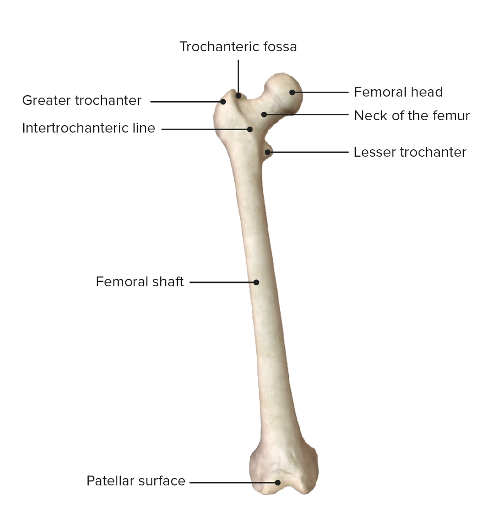
00:01 Here, we can see the main musculature on the anterior thigh. We can see that these muscles, rectus femoris, vastus medialis, vastus intermedius, and vastus lateralis, collectively known as the quadriceps. Femoris muscle lie on the anterior aspects or anterior to the femur in this right lower limb. We can see they pass down towards the tibia via this patellar ligament. So if we have a look at the origins and the insertions, then the rectus femoris is coming from the anterior inferior iliac spine. Make sure you remember those bony landmarks we pointed out. Vastus lateralis is coming from the greater trochanter and the lateral lip of the linea aspera. Vastus intermedius is coming from the anterior aspect of the shaft of the femur, whereas, vastus medialis is coming from the intertrochanteric line and the medial lip of the linea aspera. All of these muscles travel down and converge on the tibial tuberosity initially via the common quadriceps and then the patellar ligament with the patellar bone actually within the patellar ligament sitting anterior to the distal part of the femur. All of these muscles are supplied by the femoral nerve, the femoral nerve being the nerve of the anterior compartment. They’re involved in extending the leg at the knee joint. And rectus femoris is also involved in supporting iliopsoas in flexing the hip. And this is possible because rectus femoris attaches or originates from the anterior inferior iliac spine. Now we move on to look at more of these anterior thigh muscles, but we’re going to look at some muscles known as iliopsoas, and this muscle is a combination of both psoas major that’s coming down from the posterior abdominal wall, and also iliacus that lines the surface, the internal surface of the ilium. These two muscles converge into a common tendon that travels down to the lesser trochanter of the femur. And here, we’re going to look at sartorius as well. Sartorius coming down from the anterior superior iliac spine, it’s the most superficial muscle within this anterior compartment. So we have iliopsoas. 02:28 The psoas part comes from the lateral surface of T12 to L5 vertebrae, and it passes through the lesser trochanter of the femur. It’s supplied by the anterior rami of the lumbar nerves, L1 L2. Iliopsoas, the iliacus part of it, comes from the iliac crest and fossa. It joins with the tendon of psoas major and also passes through the lesser trochanter of the femur. It’s supplied by the femoral nerve. Sartorius is coming from the anterior superior iliac spine. It passes to the medial surface of the proximal tibia, and we’ll see that later on when we talk about the pes anserinus. This is also supplied by the femoral nerve. Pectineus that is coming from the superior pubic ramus and passing to the pectineal line of the femur. And we’ll look at this when we look at the adductor compartment. So there it’s in the anterior thigh. It can be easily viewed when we’re looking at the adductor compartment. We can see it briefly here, though. We’ve got pectineus coming from the superior pubic ramus, and it’s passing down onto the pectineal line, and sometimes the lesser tubercle of the femur. But you can see here how it’s closely associated with the other adductor muscle. We’ll talk about it again later. These muscles are important in doing a range of movements. So iliopsoas is important in flexing and stabilizing the hip. 04:03 Sartorius does multiple movements, and if you look at its positioning, hopefully, this makes sense. 04:08 It flexes, it abducts, and it laterally rotates the hip. It’s also involved in flexing the knee. 04:17 So if we look at sartorius, then we can see it’s running superficially, but it’s passing behind the medial condyle of the femur. So, contraction of this muscle can lead to a whole range of movement. Lateral rotation, it can flex the knee, and it’s also able to abduct the hip. Pectineus helps to adduct the femur at the hip joint. It also is a small flexor or weak flexor of the hip, and it supports medial rotation of the hip. 04:51 So make sure you’re happy with these origins, insertions, and what movements they can perform.
About the Lecture
The lecture Anterior Thigh Muscles – Anterior and Medial Thigh by James Pickering, PhD is from the course Lower Limb Anatomy [Archive].
Included Quiz Questions
Which muscle forms the lateral wall of the adductor canal?
- Vastus medialis
- Sartorius
- Vastus lateralis
- Adductor longus
- Vastus intermedius
Where does the rectus femoris originate?
- Anterior inferior iliac spine
- Anterior superior iliac spine
- Posterior superior iliac spine
- Posterior inferior iliac spine
- Anterior shaft of the femur
What is the site of origin of the rectus femoris muscle?
- Anterior inferior iliac spine
- The greater trochanter
- Anterior shaft of the femur
- Lateral lip of the linea aspera
- Medial lip of the linea aspera
Customer reviews
3,0 of 5 stars
| 5 Stars |
|
1 |
| 4 Stars |
|
0 |
| 3 Stars |
|
0 |
| 2 Stars |
|
0 |
| 1 Star |
|
1 |
idk what the below review is talking about, this is clearly an introduction-style lecture and it's been done well. does well to get cogs whirring
Anatomy is explained best with annotated images and pointers on the various parts as the lecturer speaks. Not through tabulation of facts.


