Playlist
Show Playlist
Hide Playlist
Anterior Mediastinum
-
Slides Anatomy Anterior Mediastinum.pdf
-
Reference List Anatomy.pdf
-
Download Lecture Overview
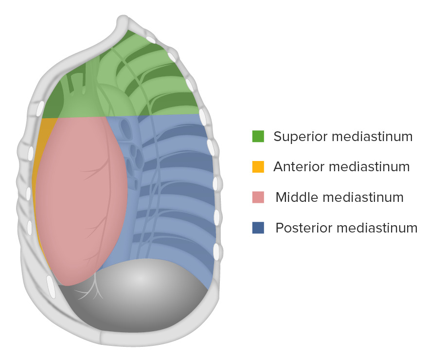
00:01 Now that we've seen the lungs, we can talk about the space between the lungs, which is called the mediastinum. 00:09 And as a space, it's sometimes hard to conceptualize a space. 00:12 So we'll start by talking about the borders of the mediastinum. 00:17 The first border superiorly is the superior thoracic aperture. 00:22 Inferiorly, we have the diaphragm. 00:26 Anteriorly, we have the sternum. 00:29 And posteriorly, we have the thoracic vertebra. 00:32 And of course, laterally, as we've defined this on either side, we have the parietal pleura, on the right and left. 00:40 The mediastinum is divided into more parts. 00:43 In order to talk about those parts, we first have to come up with an imaginary structure called the transverse plane. 00:51 So this isn't a real anatomic structure. 00:53 It's one we're going to imagine starting from the sternal angle, and going straight back to the vertebra till it hits around the T4-T5 area. 01:03 And that transverse plane is going to divide the superior mediastinum above from the inferior mediastinum below. 01:14 Now the superior mediastinum is just the superior mediastinum. 01:18 But the inferior mediastinum gets further subdivided. 01:23 And ordered to describe those subdivisions, we first have to find the middle mediastinum. 01:29 And the middle mediastinum is pretty easy. 01:32 It's defined as everything within the pericardium, which is pretty much just the heart and the origins of the great vessels that attach to the heart. 01:40 Anterior to that is the anterior mediastinum. 01:44 And posterior to that is the posterior mediastinum. 01:49 If we zoom in on that superior mediastinum a little bit more, we again see the boundaries, same as before the superior thoracic aperture, and inferiorly is that transverse plane that we're imagining starting at the sternal angle. 02:05 Anteriorly, we have the sternum. 02:07 Posteriorly, the vertebra. 02:09 And again, the parietal pleura on either side laterally. 02:13 There's some really important structures that go through here. 02:17 First of all, we have some very important nerves. 02:20 We have the vagus nerves coming through, and we have the phrenic nerves. 02:25 We also have some very prominent veins. 02:27 We have the brachiocephalic veins, coming down and joining with the superior vena cava. 02:34 We have very prominent arteries too. 02:36 We have the aortic arch and its prominent branches, the brachiocephalic trunk, the left common carotid artery, and the left subclavian artery. 02:46 All big stuff and all stuff we're gonna see in greater detail when we talk about these vessels. 02:51 But there's also a structure here that we kind of see. 02:54 We see a little bit of the thymus. 02:57 We're gonna see the rest of it when we go down into the inferior mediastinum. 03:03 Starting with the middle mediastinum because it's the easiest. 03:06 The middle mediastinum is anything within the pericardium. 03:10 And there's not much besides the heart and the origins of the great vessels that attached to it. 03:16 We're gonna have a separate section on those don't worry. 03:19 Anterior to the pericardium is the anterior mediastinum. 03:25 And it's pretty small. 03:27 Its boundaries are the transverse plane superiorly. 03:31 The diaphragm, inferiorly. Sternum, anteriorly. 03:36 And that pericardium, posteriorly. 03:38 And of course, on either side, we have the parietal pleura. 03:42 But it's a pretty small space. 03:45 And essentially, the only thing we have in here that we have to worry about is the rest of that thing that we saw in the superior mediastinum a little bit called the thymus. 03:53 And how much we actually see depends on the age of the person. 03:59 And I'll explain that at the very end of this section. 04:03 First, let's cover the posterior mediastinum. 04:06 So the posterior mediastinum is everything posterior to the middle mediastinum. 04:10 Therefore, everything posterior to the pericardium. 04:14 And its superior border is again the transverse plane, inferior border is the diaphragm, anterior is the pericardium, posterior the thoracic vertebra, and on either side we have the parietal pleura. 04:28 Now, the posterior mediastinum is quite a bit bigger. 04:30 So there's a lot more traveling through here. 04:33 For example, we have the bronchi going into the lungs. 04:37 We have the esophagus on its way down to the abdomen. 04:40 We have both the aorta and the Azygos system. 04:44 We have the sympathetic trunks on either side of the vertebra. 04:49 Before we finish, I promised I would explain what I meant by how much of the thymus you can see. 04:54 So, what is the thymus? So the thymus is a lymphoid organ that it's very important for T cell development. 05:03 But it's really important more so in the early stages of life. 05:07 It's very prominent in the anterior mediastinum in infancy and early childhood. 05:13 However, over time, it starts to regress, or what we say involute and turn to fat. 05:20 So that by adulthood even though there are some little bits of functional thymic tissue that are still in there, it becomes indistinguishable from just normal body fat.
About the Lecture
The lecture Anterior Mediastinum by Darren Salmi, MD, MS is from the course Thorax Anatomy.
Included Quiz Questions
What is the posterior border of the mediastinum?
- Thoracic vertebrae
- Sternum
- Esophagus
- Diaphragm
- Azygos vein
What is not a structure within the superior mediastinum?
- Inferior vena cava
- Phrenic nerves
- Vagus nerves
- Aortic arch
- Brachiocephalic veins
Which structure is present within the anterior mediastinum?
- Thymus
- Thyroid
- Trachea
- Aortic arch
- Cochlea
Customer reviews
5,0 of 5 stars
| 5 Stars |
|
5 |
| 4 Stars |
|
0 |
| 3 Stars |
|
0 |
| 2 Stars |
|
0 |
| 1 Star |
|
0 |


