Playlist
Show Playlist
Hide Playlist
Anterior Compartment – Anatomy of the Forearm

-
Slides 06 UpperLimbAnatomy Pickering.pdf
-
Download Lecture Overview
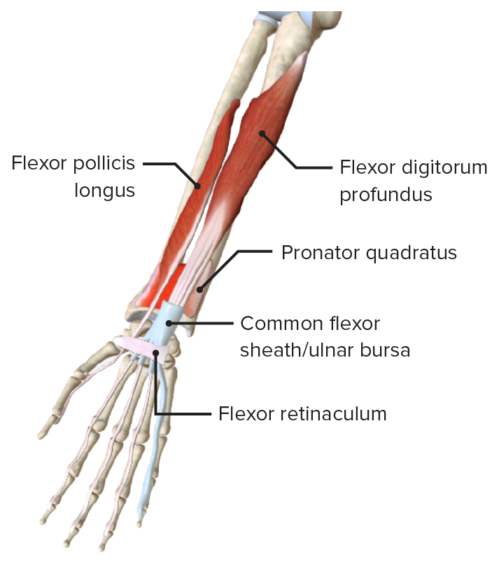
00:01 start off by looking at muscles in this anterior compartment. Now, there is a warning, there is a lot of muscles here. There are a lot of muscles in this anterior compartment of the forearm. But conveniently, they have been split up into various layers. And we can look at these layers individually and the muscles that lie within each of these three layers, a superficial, a middle, and then a deep layer. And we’ll look at these layers individually. 00:31 So first of all, we’ll start with the superficial layer. We can see here on this superficial layer we have pronator teres, we have flexor carpi radialis, we have palmaris longus, and we have flexor carpi ulnaris. These are all coming from a common origin. So they’re originating from a very similar place, this being the medial epicondyle. There can be some slight variations to where all of these muscles come from. But the main common origin is this medial epicondyle here. And we can see they’re radiating across the elbow joint and attaching to various places on the forearm and also in the hand. So we can see pronator teres is running across to the radius. We can see flexor carpi radialis is running towards the second metacarpal. We can see palmaris longus is running towards the palm of the hand. And we can see flexor carpi ulnaris here is running towards one of the carpal bones. So let’s look at this in more detail. We can see in the superficial layer, we have pronator teres, flexor carpi radialis, palmaris longus, and flexor carpi ulnaris. If we look at pronator teres, we’ve got two heads of pronator teres because they have a different origin; an ulnar head coming from the coronoid process of the ulna, and a humeral head which is coming from that medial epicondyle. This is why it’s important to have those bony attachments, those bony landmarks from the osteology lecture previously. The pronator teres muscle is going to insert onto the middle of the lateral surface of the radius. 02:20 So if we go back, we can see the pronator teres coming from the medial epicondyle. And also coming from the ulna, its ulnar head, is passing towards the shaft of the radius, we can see it here. We then remind ourselves flexor carpi radialis, palmaris longus, flexor carpi ulnaris. 02:40 And we can look at their attachments. Flexor carpi radialis and palmaris longus are coming from the medial epicondyle of the humerus. So, both of these are coming from the medial epicondyle of the humerus. As we saw, the flexor carpi radialis attaches to the base of the second metacarpal, whereas, palmaris longus passes towards the palm of the hand and attaches to what’s known as the palmar aponeurosis and the flexor retinaculum. 03:11 And we’ll look at this as we pass towards the hand in next lecture. All of these muscles, pronator teres, flexor carpi radialis, palmaris longus, are supplied by the median nerve. 03:23 The one muscle in the superficial layer that is supplied by the ulnar nerve is flexor carpi ulnaris, and this lied most laterally, and this was positioned most medially within the forearm. It originates from the olecranon and the posterior surface of the ulnar, and it passes to a range of bony landmarks, the pisiform and the hook of the hamate, also, the fifth metacarpal. So flexor carpi ulnaris is passing to these, inserting to these bony landmarks. It’s different from the other superficial muscles in this compartment, in that is innervated via the ulnar nerve. If we look at the function of these muscles, well, pronator teres, its name gives it away, it pronates the forearm. It can also because it crosses over the elbow joint, flex the forearm. Flexor carpi radialis is important at flexing the wrist. It also, because it’s on this lateral aspect, contraction can abduct the hand at the wrist joint. Palmaris longus, this flexes the hand at the wrist. It also tenses the palmar aponeurosis, the palmar aponeurosis being a tough fibrous tissue in the palm of the hand, and tensing this is important when forming a grip. Flexor carpi ulnaris is very similar to flexor carpi radialis, in that it flexes the wrist. But as it’s running down this medial side, it also adducts the wrist, or it deviates it to the ulnar side. 04:58 So now if we go back and have a look at the middle layer, then there really is only one muscle that I really want to talk about in this middle layer, and that is flexor digitorum superficialis. Flexor digitorum superficialis, as its name suggests, is a flexor of the digits. 05:19 As we call it, flexor digitorum superficialis, it means that there’s going to be a flexor digitorum profundus, and we can see that on this picture here. But we’re just concentrating on flexor digitorum superficialis. We can see that its long tendons pass all the way to the middle phalanx of the digits. We can see that here. If we have a look at the attachments of those muscles, we can see flexor digitorum superficialis. It has got two heads. 05:57 It has got a humero-ulnar head, and this head is coming from the medial epicondyle. 06:03 It’s coming from the coronoid process as well, and that’s where we have a humero-ulnar head. 06:09 It has a part coming from the humerus, the middle epicondyle, and a part coming from the ulna, the coronoid process. It also has a radial head, and it’s coming from the shaft of the radius. So the flexor digitorum is coming from a whole wide range of regions from the humerus, the ulna and the radius. This muscle belly, eventually, is going to give rise to long tendons that go and attach to the middle phalanges of the medial four digits. So, there are all the digits, but not including the thumb; so digits 2, 3, 4, and 5. It’s supplied by the median nerve. Nerve supply to flexor digitorum superficialis is the median nerve. Importantly, it flexes the hand at the wrist joint. It also flexes the proximal interphalangeal joint. That’s the joint between the proximal and the middle phalanges, the proximal interphalangeal joint. With continued action, it will flex the proximal phalanges at the metacarpophalangeal joint. So ultimately, it helps us to form a fist. 07:24 It helps us to form a fist. If we then look at the deep layer, we can see we’ve got
About the Lecture
The lecture Anterior Compartment – Anatomy of the Forearm by James Pickering, PhD is from the course Upper Limb Anatomy [Archive].
Included Quiz Questions
Which muscle inserts onto the base of the second metacarpal bone?
- Flexor carpi radialis
- Flexor carpi ulnaris
- Palmaris longus
- Pronator teres
- Flexor digitorum superficialis
Which muscle is innervated by the ulnar nerve?
- Flexor carpi ulnaris
- Flexor carpi radialis
- Flexor digitorum superficialis
- Pronator teres
- Palmaris longus
Which bone does the flexor digitorum superficialis insert onto?
- Middle phalanges of the medial 4 digits
- Proximal phalanges of the lateral 4 digits
- Proximal phalanges of the medial 4 digits
- Distal phalanges of the medial 4 digits
- Distal phalanges of the lateral 4 digits
Author of lecture Anterior Compartment – Anatomy of the Forearm

James Pickering, PhD
Customer reviews
5,0 of 5 stars
| 5 Stars |
|
1 |
| 4 Stars |
|
0 |
| 3 Stars |
|
0 |
| 2 Stars |
|
0 |
| 1 Star |
|
0 |
thnak u that perfect thnak u that perfect thnak u that perfect thnak u that perfect
