Playlist
Show Playlist
Hide Playlist
Anterior Compartment – Anatomy of the Arm

-
Slides 05 UpperLimbAnatomy Pickering.pdf
-
Download Lecture Overview
00:01
So if we look at the anterior
compartment of the arm, then they are three
muscles that I want you to be aware of in
the anterior compartment. These are coracobrachialis,
brachialis, and biceps brachii. If we start
off with coracobrachialis, this is a relatively
short muscle as its name suggests, coracobrachialis
is running from the coracoid process of the
scapula. Again this is why those bony landmarks
on the scapula were important to the shaft
of the humerus. And here we can see we have
got coracobrachialis running from the coracoid
process of the scapula to the middle third
of the humerus. It is supplied by the musculocutaneous
nerve. The musculocutaneous nerve that was
coming from the brachial plexus. This muscle
doesn’t cross the elbow joint. It just crosses
the glenohumeral joint and because it does
that it flexes and also adducts this joint.
01:04
If we go back and look at biceps brachii,
we can see on this picture. We can see biceps
brachii has two heads as its name suggests. We
have a short head that is running up towards
the coracoid process and we also have a long
head that is running up towards the supraglenoid
tubercle, the supraglenoid tubercle, that tubercle above
the glenoid cavity. The tendon of the long
head actually takes quite a long journey. It
passes up within the intertubercular groove.
01:41
The intertubercular groove forms by the greater
and lesser tubercles of the humerus. So here
we see the long head of biceps passing to
the supraglenoid tubercle by passing through
the intertubercular groove. The short head
here attaches to the coracoid process.
02:02
Here we can see the biceps which is running within
the anterior compartment of the arm, the
muscle belly of biceps and we can see its main
attachment is on to the radius, the radial
tuberosity which we can see here. It also
gives rise to the bicipital aponeurosis and that
blends with the brachial fascia. Another
muscle here is brachialis and this muscle
like biceps brachii crosses over the elbow
joints. But because it is originating from
the midshaft of the humerus it doesn't cross
the shoulder joint whereas here biceps brachii
does. Brachialis coming from the middle 3rd
of the shaft of the humerus, it passes all
the way down on to the ulnar and we can see
brachialis muscle here. This muscle is only
going to act on the elbow joint. So if you
look at biceps brachii, its long head and
short head. We can see it is originating from
the supraglenoid tubercle or the coracoid
process. It passes through the tuberosity
of the radius and it blends with the fascia
of the forearm via the bicipital aponeurosis
as that continuation of brachial fascia passes
down into antebrachial fascia. It is innervated
via the musculocutaneous nerve and it has
a whole range of functions. It is the chief supinator
of the forearm. Because of its attachment
on to the radius contraction of biceps will
actually supinate the forearm. So from this
pronated on mid prone position, the first
movement of biceps will be to supinate the
forearm. Once the forearm is supinated continued
contraction of biceps will then flex the elbow
joint. But only once the forearm is in this supinate
position will it then flex. Continued contraction
of biceps because it crosses the shoulder
joint can leads a flexion of the shoulder.
04:03
Brachialis as I mentioned comes from the distal
half of the humerus and it attaches to the coronoid
process and tuberosity of ulnar. It is also
supplied by the musculocutaneous nerve and it
flexes the elbow joint. What you notice is
that the muscles in the anterior compartment
of the arm are all supplied by the same nerve,
the musculocutaneous nerve. That terminal
branch coming from the lateral coat of the brachial
plexus is giving rise to the musculocutaneous
nerve that supplies the muscles in the anterior
compartment of the forearm. So if we look at
these in a bit more anatomical position that
actually to see these muscles we need to remove
deltoid. So in the slide at the moment we
have got a muscle here, an anterior view of
the right arm with deltoid in position. We
have also removed deltoid on this picture.
05:06
And by removing deltoid we can then see the
position of the long head of biceps here.
05:12
We can also see the short head of biceps here.
The muscle belly of biceps is in place here
and we removed it on this picture to see brachialis
lying underneath. So hopefully you could appreciate
that brachialis is sitting directly deep to
biceps brachii muscle. Its very difficult
to see brachialis muscle with biceps brachii
still in place. Just see a small bit of it
here and here. We can see what we have got
the bicipital aponeurosis passing down on to
the antebrachial fascia of the forearm. So
that we can see if we look at these notes which
we have here, that running over the intertubercular
groove, we have also noticed the transverse
humeral ligament. And that runs over the two tubercles
on the head of the humerus and this creates
a tunnel for the long head to pass through.
As I mentioned distally, the two heads of
biceps unite and this continues down as the
bicipital aponeurosis. Deep to biceps brachii
is brachialis and medial to biceps brachii
is coracobrachialis which we can see here.
06:26
So we can see in these pictures all of the muscular
put together into one anterior compartment
of the arm. If you look at the neurovascular
relations of the anterior compartment then
we can see in the diagram we have got a brachial
plexus again. We can also see we have got
some important blood vessels and then neighboring
nerves as they descend distally towards the
elbow joint. So we can see the muscular cutaneous
nerve once again. We can see the median nerve.
07:03
We can see the ulnar nerve and these are running
down this medial aspect of the arm. We can see
they are running all the way down to medial
aspect. We can see actually here the medial
intermuscular septum of the arm. I remember
that separates the anterior compartment from
the posterior compartment. Here we can pick
up the axillary artery but we can also see
its relationship to the brachial plexus and then
as it runs down it becomes the brachial artery.
07:34
And we can see the brachial artery is running in a
groove between biceps brachii and brachialis
muscle. So here you got biceps and deep to
biceps we have got brachialis muscle just
here and in the groove between we can see
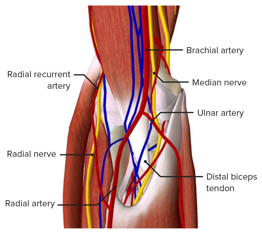
the brachial artery. We can also observe if
we go towards the anterior aspects of
the elbow, the bicipital tendon which we can
see here and that is forming a roof over the
brachial artery and the median nerve.
08:05
The brachial artery and the median nerve are running
underneath the bicipital aponeurosis and the
biceps tendon. And this is the cubital fossa and
we will cover that in future slides.
About the Lecture
The lecture Anterior Compartment – Anatomy of the Arm by James Pickering, PhD is from the course Upper Limb Anatomy [Archive].
Included Quiz Questions
Which movements does the coracobrachialis perform?
- Flexion and adduction
- Flexion and abduction
- Extension and abduction
- Extension and adduction
- Lateral rotation
Which muscle is the chief supinator of the forearm?
- Biceps brachii
- Coracobrachialis
- Triceps brachii
- Quadriceps
- Deltoid
Which nerve innervates the muscles in the anterior compartment of the arm?
- Musculocutaneous
- Median
- Axillary
- Radial
- Ulnar
Which muscle lies medially and posteriorly to the biceps brachii muscle?
- Coracobrachialis
- Brachialis
- Triceps brachii
- Deltoid
- Brachioradialis
The brachial artery runs between which 2 muscles in the lower one-third of the arm?
- Biceps brachii and brachialis
- Biceps brachii and triceps brachii
- Triceps brachii and coracobrachialis
- Coracobrachialis and brachioradialis
- Coracobrachialis and brachialis
Author of lecture Anterior Compartment – Anatomy of the Arm

James Pickering, PhD
Customer reviews
5,0 of 5 stars
| 5 Stars |
|
1 |
| 4 Stars |
|
0 |
| 3 Stars |
|
0 |
| 2 Stars |
|
0 |
| 1 Star |
|
0 |
Fantastic explanation about the topic, easy way to learn anatomy.
