Playlist
Show Playlist
Hide Playlist
Anterior Axioappendicular Muscles – Anatomy of the Shoulder

-
Slides 03 UpperLimbAnatomy Pickering.pdf
-
Download Lecture Overview
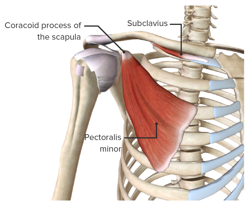
00:01 In his lecture we're going to look at the shoulder region, specifically the axioappendicular and the scapulohumeral muscles. 00:10 So we're going to look at the shoulder joints and its range of movements, and specifically how the muscles enable the shoulder to move. 00:19 The anterior and posterior axioappendicular muscles. We'll then look at the scapulohumeral muscles and how some of these form the rotator cuff coming from the scapula to the humerus. 00:33 We'll then look at the functional anatomy of these with their origin, insertion, and the movements that they can allow the shoulder to perform. 00:42 So the anterior axioappendicular muscles -- what do I mean by axioappendicular muscles? Well, these are muscles that run from the axial skeleton to the appendicular skeleton. 00:57 So they run from the axial skeleton -- the sternum, the ribs, the vertebral column to the appendicular skeleton which in this case is the upper limb, the superior appendicular skeleton -- the clavicle, the scapula, the humerus. 01:14 And here we can see the anterior muscles; we have a whole series of them. 01:18 We have pectoralis major, we can see here. 01:22 We can have pectoralis minor, we can see here. 01:25 We've got subclavius, we can see here. 01:28 And also we've got serratus anterior -- we'll come back to that later on. 01:33 What we can see is the pectoralis major muscle has a number of heads -- really it's got two heads. 01:40 It's got a clavicular head, where it's coming from the clavicle, but the axioappendicular part of it is where it's coming from the sternum and the costal margins of the ribs. 01:53 We can see this muscle, this fan-shaped muscle is running down towards the shaft of the humerus and we'll see where it attaches on the shaft of the humerus just within the intertubercular sulcus, but here not up in between the greater and lesser tubercles, quite distally down on to the shaft of the humerus. 02:14 So we got pectoralis major here. 02:17 This is a large fan-shaped muscle that's easily visible once the skin of the anterior chest wall has been reflected. 02:25 Clavicular head coming from the clavicle, and the sternocostal head coming really from the sternum and the costal cartilages. 02:34 Reflect pectoralis major and we find we have pectoralis minor. 02:39 We can see pectoralis minor here coming from ribs 3, 4, and 5, we have pectorals minor running up to the coracoid process of the scapula. 02:51 So here it's now important to remember those bony features I was talking about in the first lecture. 02:57 We can see we've got pectoralis minor running up towards the coracoid process and that lies deep to pectoralis major. 03:06 Running underneath the clavicle, running from the first rib to that groove for subclavius that I spoke about again in the first lecture on the inferior surface of the clavicle, we've got subclavius muscle we can see here, running from the sternal aspect of the first rib all the way over to the clavicle. 03:30 And here we can see these anterior axioappendicular muscles. 03:34 We'll cover serratus anterior in a moment. 03:38 We can see that the pectoralis major muscle as I mentioned it has two heads and this table really is going to detail their origin and insertion. 03:48 It's then going to look at the nerve supply and we can cover some functional anatomy here. 03:53 But here, we have the pectoralis major; we've got the clavicular head and the sternocostal head. 03:59 We can see where they're attaching as I mentioned previously. 04:03 And these are going to insert into the intertubercular sulcus specifically the lateral lip of it, but not between the two tubercles. 04:12 The nerve supply to this muscle is via the lateral and medial pectoral nerves and these are branches that are coming from the brachial plexus which we'll see. 04:23 Here we've got the specific root values, so the specific spinal cord segments that gives rise to those nerves. 04:33 The function of pectoralis major is it adducts and medially rotates the shoulder joint so it adducts, it brings the arm back against the thoracic cavity and it's also involved in medially rotating the shoulder joint. 04:52 It also pulls the scapula anteriorly so it protracts the pectoral girdle and it pulls inferiorly as well. 05:01 So pectoralis major, remember here we can see pectoralis major muscle. 05:06 Now we've got pectoralis minor, we can see pectoralis minor like I said coming from ribs 3-5, passing towards the coracoid process of the scapula and this is innervated by the medial pectoral nerves and again we can see this is coming from C8, T1 spinal cord segment. 05:26 Pectoralis minor is important in stabilizing the scapula so it holds the scapula against the posterior chest wall and it also pulls it anteriorly and inferiorly, a similar function to pectoralis major. 05:42 If we remind ourselves of the subclavius positioned inferior to the clavicle, we can see that it's running from the first rib and the sternum, the junction of those two to the middle third of the clavicle supplied by the subclavian nerve. 05:58 It stabilizes and depresses the clavicle. 06:02 We can appreciate that if we see it here coming from the junction of the sternum and the first rib, this going to be a solid, its origin, this is not going to move. 06:12 And as its muscles contract it is going to depress the clavicle. 06:16 The final muscle I want to talk about is serratus anterior. 06:21 And this is somewhat different from the previous muscles I spoke about that were coming from this more anterior aspect of the chest wall. 06:29 Serratus anterior is coming from this lateral aspect of the chest wall and it's passing backwards towards the medial border of the scapula. 06:40 So here we can see the medial border of the scapula and this is the lateral border, and this is serratus anterior muscle it's got this nice serrated edge which gives its name. 06:51 Coming from the ribs here, we can see rib two, three, four, five, six, seven, eight, nine running all the way along the chest wall. 07:02 We can see it's running between the chest wall and the scapula so this impression here is just a shading through the scapula. 07:11 It runs between the scapula and the chest wall to attach to the medial surface. 07:16 So we can see here serratus anterior coming from the external surface of the ribs 1-8. 07:23 We can see we have the fibers passing to the medial border of the scapula, see it passing in between the scapula and the posterior chest wall. 07:36 It is innervated via the long thoracic nerve and the long thoracic nerve is going to be running down in this direction. 07:44 So we can see the long thoracic nerve running down in this direction. 07:48 It's involved in protracting the scapula so it can pull the scapula forward and hold it against the posterior thoracic wall. 07:57 This is important if you are to say push off from a wall. 08:00 If you lean against a wall and pushing off, then you need the scapula to be anchored against the posterior chest wall. 08:08 If that's anchored against the posterior chest wall, then you'll be able to move backwards. 08:13 If it isn't, if there's damage say to the long thoracic nerve, then this serratus anterior cannot contract, it cannot hold the scapula against the posterior thoracic wall and then when you push off, the scapula actually pushes out into the skin of the back and you have something called winged scapula. 08:34 So damage to the thoracic nerve via trauma or knife attack into the axilla or breast surgery or removal of lymph nodes for cancer treatment can damage the long thoracic nerve and lead to winged scapula. 08:47 If we have to look at these at a more anatomical arrangement, then here we can see we've got pectoralis major here, we can see we got serratus anterior sitting underneath it here, we can see that the fan shaped pectoralis major is passing towards the arm, and we've got this muscle here, deltoid, which we'll cover in a few moments time. 09:12 But what we can see is that between pectoralis major here and deltoid, we have this deltopectoral triangle, and that triangle is important as it receives the cephalic vein. 09:25 As we have mentioned in the previous lecture, the cephalic vein passing up towards the axillary vein runs in the deltopectoral triangle and here we have the formation of the deltopectoral triangle. 09:39 So here we can see pectoralis major and we could see serratus anterior. 09:43 If we reflect pectoralis major over here, on this side of the screen, we can see here the reflected cartilage of pectoralis major, we can see the pectoralis major against the lateral lip of the humerus, intertubercular sulcus, and we can see pectoralis minor. 10:02 What we can see directly beneath pectoralis minor is a blood vessel here and another blood vessel here and a yellow nerve. 10:11 These indicate the axillary artery, the axillary vein, and the brachial plexus; and these are running deep within the axilla running deep to pectoralis minor -- we can see them running along here, and we'll look at these in the next lecture. 10:27 So here we can see pectoralis major, we can see once it's reflected, we can see pectoralis minor and here we can see some of the digits of serratus anterior.
About the Lecture
The lecture Anterior Axioappendicular Muscles – Anatomy of the Shoulder by James Pickering, PhD is from the course Upper Limb Anatomy [Archive].
Included Quiz Questions
Which statements describe the pectoralis major muscle? Select all that apply.
- It has a clavicular head.
- Its origin is from the 2nd and 3rd ribs.
- It has a sternocostal head.
- It is a fan-shaped muscle.
- It is attached to the lateral lip of the bicipital groove.
Which statement describes the origin of the pectoralis minor muscle?
- It arises from the 3rd, 4th, and 5th ribs.
- It arises from the 1st rib.
- It arises from the coracoid process.
- It arises from the sternum.
- It arises from the humerus.
Which function is performed by the pectoralis major muscle?
- Adducts and medially rotates the shoulder joint
- Adducts and laterally rotates the shoulder joint
- Abducts and laterally rotates the shoulder joint
- Abducts and medially rotates the shoulder joint
- Pulls the scapula inferiorly
Which muscle is supplied by the long thoracic nerve?
- Serratus anterior
- Subclavius
- Pectoralis major
- Pectoralis minor
- External intercostal muscles
Which nerve roots are injured if the result is winging of the scapula?
- C5-C7
- T1-T4
- C8-T5
- T2-T8
- C4-C5
Which vein runs in the deltopectoral triangle?
- Cephalic vein
- Basilic vein
- Superior vena cava
- Jugular vein
- Axillary vein
Which group is the nerve supply of the sternocostal head of the pectoralis major muscle?
- C7, C8, and T1
- C1 and C2
- C3 and C4
- C4 and C5
- C6 and C7
Author of lecture Anterior Axioappendicular Muscles – Anatomy of the Shoulder

James Pickering, PhD
Customer reviews
5,0 of 5 stars
| 5 Stars |
|
2 |
| 4 Stars |
|
0 |
| 3 Stars |
|
0 |
| 2 Stars |
|
0 |
| 1 Star |
|
0 |
So well organized and clearly explained! The way you organize the material makes anatomy reasonable... the puzzle pieces fit.
I am really enjoying this, helps me understand better and it makes it possible to review after I have had a lecture at my university or even before.
