Playlist
Show Playlist
Hide Playlist
Anterior and Posterior Arteries of the Cerebral Cortex

-
Slides 17 CerebralCortex BrainAndNervousSystem.pdf
-
Reference List Anatomy.pdf
-
Download Lecture Overview
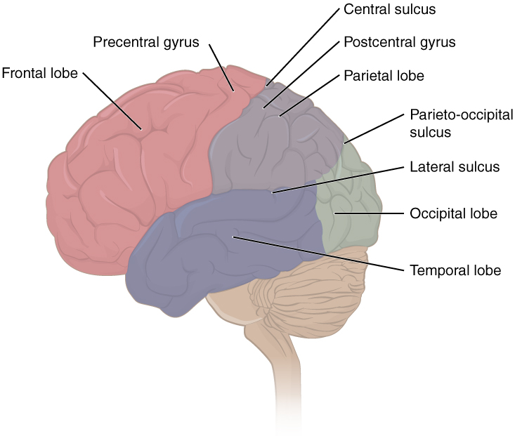
00:00 Taking a look at our arteries that supply the cerebrum specifically, we’re going to take a look here on this medial view the course of the anterior cerebral artery. So again, this is going to travel on the medial side of the cerebral cortex as an anterior course passing anterior to your corpus callosum and then circles back, if you will and winds back over the corpus callosum. Then it will send some branches over the superior aspect of the cerebral cortex that it supplies. The posterior cerebral artery is shown in through here as a much more posterior course as you would imagine. You can see it branching. 00:54 You can see it supplying the more posterior aspects of the cerebrum. Now, I want you to focus on each one of these arteries in a little greater detail. What we want to do is to understand the actual distribution of first the anterior cerebral artery and then the posterior cerebral artery. So, the area in blue on this lateral view represents the territories supplied by the anterior cerebral artery. So those areas would include the medial, frontal, and parietal lobes, so more on the medial side of this view. The anterior cerebral artery also supplies the anterior limb of the internal capsule. It also will supply most of the corpus callosum, the structure that connects the cerebral hemispheres to one another. 01:57 Here we see on the medial view in blue again, shaded in blue the distribution of the anterior cerebral artery. All the medial side of the frontal lobe, all the medial side of the parietal lobe essentially is being supplied here. Now, we want to take a look at the middle cerebral artery and the territory that it supplies. That’s the area that’s shown in through here. The territory supplied by your middle cerebral would be the lateral frontal, lateral parietal lobes, as well as a portion of the temporal lobes. With respect to the internal capsule, it’s going to supply the genu and its posterior limb. This is the vessel that will give rise to branches that supply most of the structures that belong to the basal ganglia. Posterior cerebral artery is next. We can see the distribution on the lateral side is somewhat minimal, more so on the medial side. It will supply the occipital lobe which we see here. It supplies this portion of the temporal lobe laterally. It’s also responsible for the arterial supply to the midbrain which would be a structure outside of the cerebrum or the cerebral cortex obviously. That would be a brainstem structure. 03:56 Arterial supply of the posterior cerebral artery here in the medial view, we can see it supplying the occipital lobe. Then we see it supplying this area of the temporal lobe. 04:10 This particular image allows us to understand the arterial supply to the homunculus. 04:16 This is very useful for you clinically in establishing where a stroke may be. So if there’s paralysis of the lower limb for example versus paralysis of the upper limb, this will allow you to identify which artery is involved in producing these symptoms. 04:39 So if we take a look here, we have our famous homunculus. Here is the abdominal area, thoracic area. We see the upper limb and a large area for the hand. Of course, our face is out here and our larynx is here. Then on the medial side, we see the lower extremity with the feet being the most inferiorly located here on the medial side of the precentral gyrus. 05:08 This area shaded in kind of the bluish color is going to be the supply to the homunculus coming from the anterior cerebral artery. These areas here on either side shaded in the red color are going to be the areas of the homunculus supplied by the middle cerebral artery. 05:32 So, let’s say for example that you have a patient who has paralysis of the lower extremity. 05:41 You’re trying to isolate which artery would be involved in producing that paralysis and understanding the blood supply here to the lower limb. Within the precentral gyrus, this is going to involve the anterior cerebral artery. Next, arterial supply to the internal capsule, the internal capsule has an anterior limb that we see running within here. 06:11 It has a band called the genu right in through here. Then we see at that band the internal capsule kind of changes its course and then has this orientation or direction. This is the posterior limb. What’s important about the internal capsule is there is a somatotopic organization with regards to the muscles that are being supplied through nerve fibers traveling in the internal capsule. So, we can see here at the genu area, you’re looking at muscles that are associated with the face and neck. Then as we proceed down the posterior limb of the internal capsule, as we go in this direction, you’re moving down the direction of this homunculus within the internal capsule. So that by the time you get here, you're looking at the feet area. So if you think about a stroke and what is supplied by the cerebral arteries here, the anterior cerebral artery supplies the anterior limb. So, a stroke there would have no impact on muscle control. The genu in the posterior limbs, however are supplied by the middle cerebral artery. So there is potential that if it’s involved in a stroke that you could have a lesion within the internal capsule as a result. 07:50 Lastly, I want you to understand watershed territory. Watershed area is an area of the brain that’s fed from two arterial sources. So, this could be between anterior cerebral artery, middle cerebral artery for example or it could be between the middle cerebral, posterior cerebral as well. These are just a couple of watershed possibilities. If we take a look here again, this was an image that we used before to identify the territories supplied by your anterior cerebral artery, that in blue and then the middle cerebral artery is this area shown in through here. So, if you focus your attention here in the frontal lobe, you will see this boundary between the anterior cerebral and the middle cerebral. So there’s an overlap of those two blood vessels to this area of cortex. So, a watershed territory here would be fed by the anterior cerebral and the middle cerebral arteries. You can have what is known as a watershed infarct if the driving pressure to this watershed area is diminished. 09:14 Another example is that you certainly could have a watershed territory between the middle cerebral artery and the posterior cerebral artery. Here is the territory supplied by your posterior cerebral artery. Here is the territory supplied by the middle cerebral artery. 09:33 So this would be a watershed area, for example, here between the posterior cerebral and the middle cerebral arteries.
About the Lecture
The lecture Anterior and Posterior Arteries of the Cerebral Cortex by Craig Canby, PhD is from the course Cerebral Cortex. It contains the following chapters:
- Anterior and Posterior Arteries
- Arterial Supply of the Homunculus
Included Quiz Questions
Which of the following is NOT supplied by the anterior cerebral artery?
- Motor and sensory cortex of upper limb and face
- Medial frontal lobe
- Major part of the corpus callosum
- Anterior limb of the internal capsule
- Parietal lobe
Which of the following supplies the lateral part of the frontal lobe?
- Middle cerebral artery
- Anterior cerebral artery
- Medial cerebral artery
- Lateral cerebral artery
- Posterior cerebral artery
Which of the following statements about the internal capsule is true?
- The genu of the internal capsule is supplied by the middle cerebral artery.
- The anterior limb of the internal capsule is supplied by the middle cerebral artery.
- The anterior limb of the internal capsule is supplied by the posterior cerebral artery.
- The genu of the internal capsule is supplied by the anterior cerebral artery.
- The posterior limb of the internal capsule is supplied by the posterior cerebral artery.
If the right leg is paralyzed, which of the following vascular territories is most likely to have an infarction?
- Anterior cerebral artery
- Superior division of middle cerebral artery
- External carotid artery
- Posterior cerebral artery
Which of the following statements about the watershed area is true?
- This area is susceptible to infarction if the driving pressure is diminished.
- This area is highly resistant to ischemia.
- This area is oxygenated by veins.
- This area is supplied by a single artery.
- This area is highly perfused.
Author of lecture Anterior and Posterior Arteries of the Cerebral Cortex

Craig Canby, PhD
Customer reviews
5,0 of 5 stars
| 5 Stars |
|
1 |
| 4 Stars |
|
0 |
| 3 Stars |
|
0 |
| 2 Stars |
|
0 |
| 1 Star |
|
0 |
A series of very useful and didactic lessons, amazing! Thank you so much Dr. Canby.
