Playlist
Show Playlist
Hide Playlist
Anatomy of the Uterine (Fallopian) Tubes

-
Slides Anatomy of the Uterine (Fallopian) Tubes.pdf
-
Reference List Anatomy.pdf
-
Download Lecture Overview
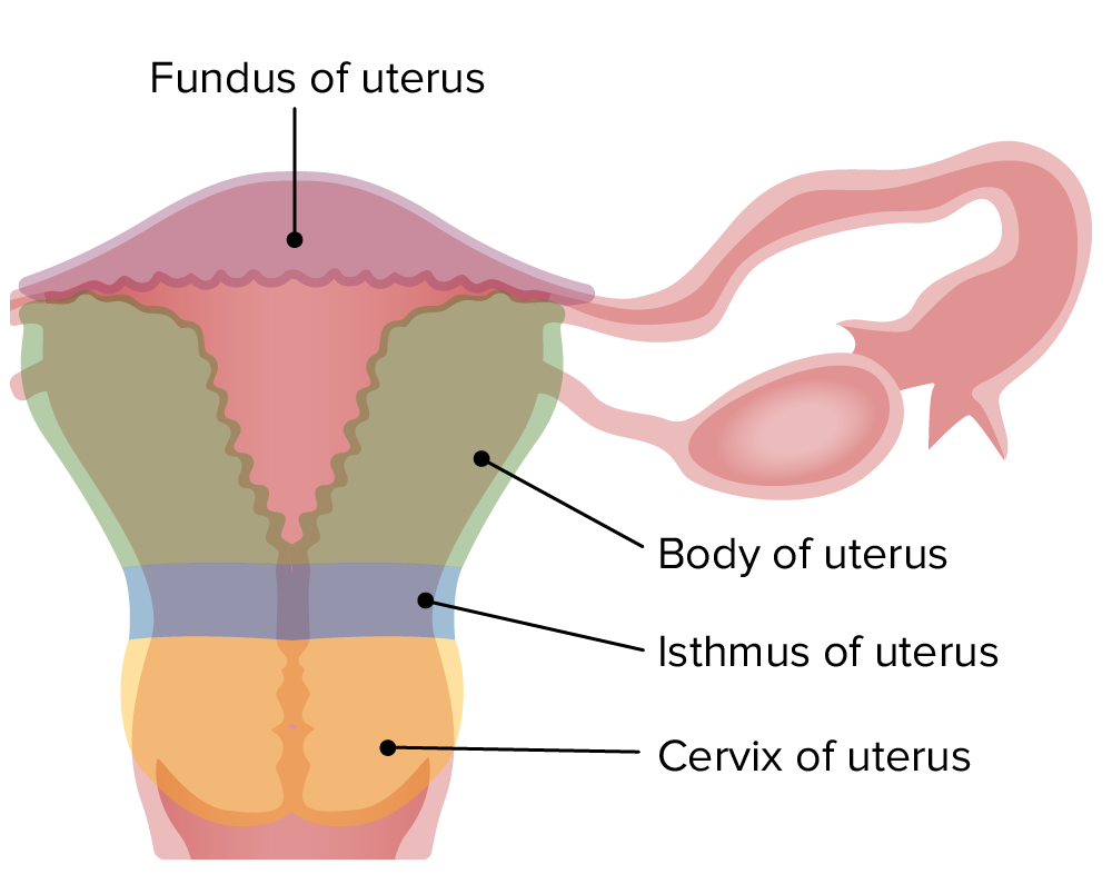
00:01 Now let's turn our attention to the uterine tubes. 00:06 So we have a pair of uterine tubes that emerge from the uterine horns laterally from the bladder. 00:12 So we can see here we have the uterus. 00:15 We've got the corner of the uterus, which is the beginning of those uterine tubes. 00:19 So we have the uterine part of those tubes. 00:22 It then extends laterally as the isthmus. 00:24 We then have the dilation of the uterine tube as the ampulla. 00:28 And then here we can see where you usually have the site of fertilization. 00:33 So that's where a sperm has passed into the cavity of the uterus. 00:37 It's migrated to one of these uterine tubes, and here is usually where that sperm will come into contact with an oocytes, an egg, that's been released via the ovary. 00:47 We can have the infundibulum, which is this large finger like structure that gives rise to these fimbriae that during ovulation will help to collect an egg that's passed out of the ovary and enter it into the uterine tube. 01:00 So here we can see the ovary in position. 01:03 All of the uterine tubes is intraperitoneal. 01:07 And we can see and appreciate how the uterine tube is intraperitoneal when we look at the broad ligament in a few slides time.
About the Lecture
The lecture Anatomy of the Uterine (Fallopian) Tubes by James Pickering, PhD is from the course Anatomy of the Female Reproductive System.
Included Quiz Questions
What is the usual site of fertilization?
- Ampulla
- Isthmus
- Uterus
- Cornu of uterus
- Infundibulum
Author of lecture Anatomy of the Uterine (Fallopian) Tubes

James Pickering, PhD
Customer reviews
5,0 of 5 stars
| 5 Stars |
|
5 |
| 4 Stars |
|
0 |
| 3 Stars |
|
0 |
| 2 Stars |
|
0 |
| 1 Star |
|
0 |
