Playlist
Show Playlist
Hide Playlist
Anatomy of the Pancreas and Biliary Tree

-
Slides Anatomy of the Pancreas and Biliary Tree.pdf
-
Reference List Anatomy.pdf
-
Download Lecture Overview
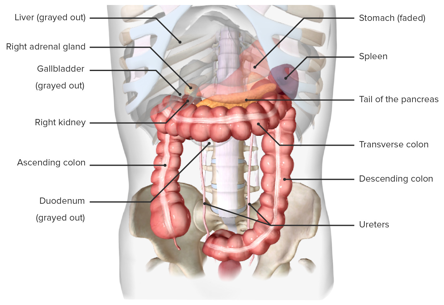
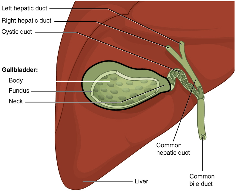
00:01 So now, let's move on to the Biliary Tree. 00:04 We touched this when we looked on the liver. 00:06 We talked about the liver in a previous video. 00:09 But now, let's have a look at it again within the context of the pancreas, and various structures associated with the pancreas, and how they form into this structure, this complex passageway of various channels. 00:21 So here we have the gallbladder. 00:23 And we can remind ourselves that we have the fundus of the gallbladder. 00:26 The part that's poking out on the inferior border of the liver. 00:29 We have the main body before it tapers into a neck, and then the cystic duct. 00:34 The cystic duct is connecting the gallbladder to the biliary tree. 00:39 Here we have the right hepatic duct and the left hepatic duct, which are coming from the two functional lobes of the liver, the left and right respectively. 00:47 And they give rise to the common hepatic duct And the union of the common hepatic duct and the cystic duct, we have the common bile duct. 00:56 And the common bile duct, we can see here is now passing through the free edge of the lesser omentum, the free margin of the lesser omentum, which we can see here. 01:06 And it's running alongside the hepatic artery proper, and also the portal vein. 01:11 So one of three structures in the portal triad that's running along the free edge of the lesser omentum. 01:18 If we then have a look, we can see the superior part of the duodenum. 01:22 And as it passes posterior to the superior part of the duodenum, it runs into the substance of the pancreas. 01:29 So here, we can see the bile duct running into the substance of the pancreas, where it's going to be met by the pancreatic duct. 01:37 The pancreatic duct runs along the entire length of the pancreas, starting from the tail, running through the neck, and running through the body, and then the neck of the pancreas where it then passes through the head and it unites with the bile duct. 01:53 So here the main pancreatic duct unites with the bile duct and it forms this large dilation which we can see here. 02:01 So this dilation is going to be called the major duodenal papilla which opens up into the duodenum. 02:09 The dilation is that hepatopancreatic ampulla. 02:13 So here, we can see the hepatopancreatic ampulla which is being fed into via the bile duct coming from the liver, and the main pancreatic duct, which is coursing along the path of the pancreas. 02:27 The hepatopancreatic ampulla, often called the ampulla of Vater in many textbooks, opens up into the duodenum via the major duodenal papilla. 02:38 Surrounding this papilla, surrounding this opening is the sphincter of the hepatopancreatic ampulla. 02:46 And again, some textbooks may call this the sphincter of Oddi. 02:49 But it's often helpful to use the actual anatomical name because it helps you work out what it is the hepatopancreatic ampulla or sphincter. 02:58 Ampulla is dilation of the formation of the hepatopancreatic, the bile duct, and the main pancreatic duct. 03:06 We also have an Accessory pancreatic duct. 03:09 This doesn't receive anything from the bile duct. 03:12 It's purely an additional pancreatic duct taking pancreatic juices into the duodenum. 03:18 These juices pass through the accessory pancreatic duct into the duodenum via the minor duodenal papilla. 03:24 And this is situated superior to the major duodenal papilla. 03:28 It can be quite variable. Not everyone has one. 03:30 And if you do, it can be quite difficult to see.
About the Lecture
The lecture Anatomy of the Pancreas and Biliary Tree by James Pickering, PhD is from the course Anatomy of the Pancreas and Spleen.
Included Quiz Questions
Which statement regarding the minor duodenal papilla is accurate?
- It is the opening of the accessory pancreatic duct.
- It has a sphincter that controls its activity.
- It is located inferior to the major duodenal papilla.
- The bile duct opens here.
- The main pancreatic duct opens here.
Which cells in the pancreas produce pancreatic enzymes?
- Acinar cells
- Alpha cells
- Beta cells
- Chief cells
- Enterochromaffin cells
What is the function of the accessory pancreatic duct?
- It drains the head of the pancreas and the uncinate process.
- It drains the tail of the pancreas.
- It produces pancreatic enzymes.
- It helps in the drainage of bile coming from the gallbladder.
- Stones are usually formed here.
Author of lecture Anatomy of the Pancreas and Biliary Tree

James Pickering, PhD
Customer reviews
5,0 of 5 stars
| 5 Stars |
|
5 |
| 4 Stars |
|
0 |
| 3 Stars |
|
0 |
| 2 Stars |
|
0 |
| 1 Star |
|
0 |
