Playlist
Show Playlist
Hide Playlist
Anatomy of the Ovaries

-
Slides Anatomy of the Ovaries.pdf
-
Reference List Anatomy.pdf
-
Download Lecture Overview
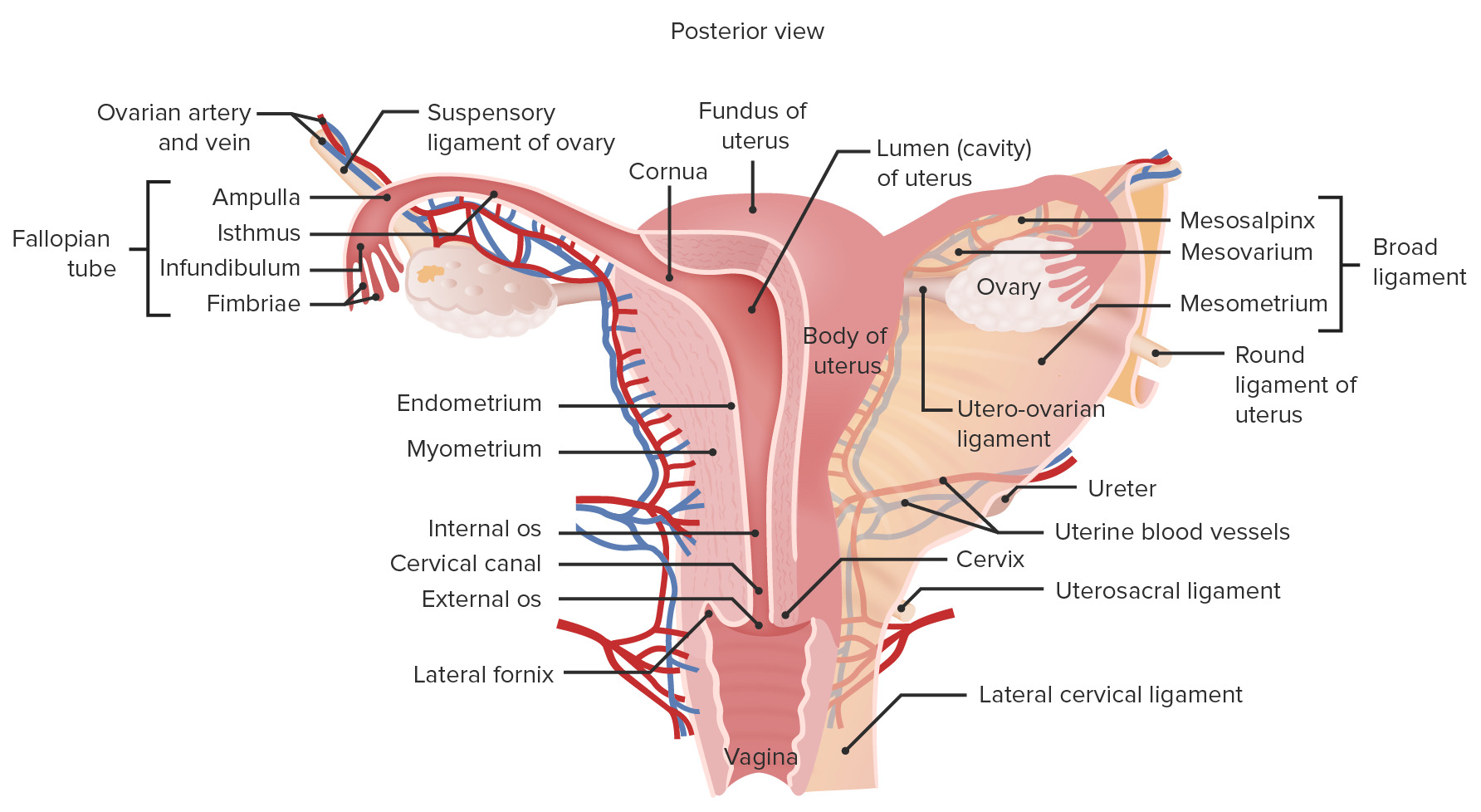
00:01 First of all, let's have a look at the ovaries. 00:05 So here again, we're looking into the pelvis. 00:08 A slightly different position here, because we're looking into a left pelvis. 00:14 So we can see the bottom right hand side of the screen, we have the pubic symphysis again. 00:19 And then most posteriorly, top left we have the sacrum. 00:23 So here we can see the bladder and the uterus just to zoom out. 00:27 And this space between these blood vessels, and what we'll soon see as the ureter is known as the ovarian fossa. 00:34 So this region indicated here is where the ovary will be located. 00:38 Superiorly, we'll find the external iliac artery and vein. 00:42 So, superior, also anterior to the ovary will see these blood vessels. 00:48 Inferiorly, we'll see the obturator artery and vein. 00:51 Notice the nerve isn't included in this split space. 00:54 So, posteriorly, we can see we have the internal iliac vein and the ureter, in this region here. 01:01 We can also include the internal iliac artery as a posterior boundary. 01:06 And then more anteriorly and slightly inferiorly we'll see we have the broad ligament of the uterus. 01:12 So the ovary position within that ovarian fossa is surrounded by a number of structures. 01:18 Superiorly, external iliac artery, external iliac vein, and a kind of superior anterior position. 01:25 Anteroinferiorly of the broad ligament of the uterus. 01:28 And then posteriorly, we have a number of blood vessels in the ureter. 01:32 And then inferiorly we have the obturator artery and vein. 01:36 A key structure that we're missing in the moment though, is the obturator nerve. 01:40 And the obturator nerve lies lateral to the ovary. 01:44 So the obturator nerve is compressed by the ovary medially, and the pelvic wall laterally. 01:50 And increase in size of the ovary, so hyperplasia of the ovary, for example, may cause compression of the obturator nerve, which will lead to a deficit within the medial compartment of the leg. 02:04 Let's continue looking at the ovary in relation to the uterine tube. 02:08 So here we can see the uterus. 02:10 We can see the uterine tube passing out from the uterus towards the ovary. 02:14 And here we can see the fimbriae, these fingerlike projections coming from the uterine tube to associate themselves with the ovaries on either side. 02:24 The superior pole of the ovary is connected to the posterior abdominal wall via the suspensory ligaments, and that has an important blood vessel running through it to supply the ovary, the ovarian artery. 02:35 And the inferior pole here is in connection to the bladder. 02:38 Not in a way to transmit any substance, like the sperm or the oocytes coming from the ovary, but as a way to support the ovary in position to hold it in place. We have the ovarian ligament. 02:51 And this is a thickening of peritoneum forming the broad ligament. 02:54 Again, we'll come to in a moment or two. 02:57 Here we have the medial surface and the lateral surface of the ovaries in position here. 03:03 So now let's have a look at the ligaments of the pelvis which we spoke about. 03:07 And importantly, these are collectively within what's known as the broad ligament. 03:12 Now, this essentially, is a double layer of peritoneum that is emerging lateral to the body of the uterus. 03:20 We can see it has a thickening anteriorly, which is the round ligament. 03:25 And this will follow a similar path to that of the ductus deferens in the male. 03:29 So the round ligament is a thickening of the broad ligament and it passes through the inguinal canal. 03:35 So it runs through the deep inguinal ring, the inguinal canal, and then the superficial inguinal ring to blend with the substance on the mons pubis. 03:44 The fatty tissue around the opening to the vagina. 03:47 So the mons pubis and the various labia majora around the lateral aspects of the vaginal opening. 03:54 So the round ligament is another important structure that helps to hold the uterus in place. 04:00 Another one that runs directly anterior from the cervix to the pubic bone is the pubocervical ligament. 04:06 And we have a similar one that runs posteriorly, and this is the cardinal ligament. 04:10 And these ligaments are important in holding the uterus in place. 04:15 Essentially, the uterus is just wedged between the bladder anteriorly and the rectum posteriorly. 04:20 And to help hold it in position to support it, we have a number of these ligaments. 04:26 A final ligament, the uterosacral ligament runs posteriorly deep to the cardinal ligament. 04:31 It's running posteriorly from the uterus to the sacrum as its name indicates. 04:37 So all of these supportive structures are kind of wrapped up within what we know is the broad ligament. 04:43 So what is the broad ligament? Essentially, the broad ligament is a peritoneal ligament. 04:49 And if you remember from the peritoneal topic, a peritoneal ligament is a double layer of peritoneum. 04:57 So what's happened here is the peritoneum is within the pelvic cavity it's run over the bladder, and then run over the uterus to run over the rectum. 05:06 But as it runs over the uterus, where it emerges laterally and the uterus stops those two layers. 05:14 So the layer that was on the anterior aspect of the uterus, and the layer those on the posterior aspect of the uterus. 05:21 As they emerge laterally, they just unite together and they come together as a double layer of peritoneum, and that is the broad ligament. 05:31 So, it's literally, as if I am the uterus, and my arms outstretched are the uterine tubes and you were to place a tablecloth over my head, then here where the tablecloth is positioned, those two layers come together, lateral to the uterus. 05:47 So they come together lateral to the uterus. 05:49 This is our broad ligament. 05:51 And we have a number of structures that run within that double layer of peritoneum. 05:56 They run within the broad ligament and they can become thickened. 06:01 So here we can see the uterus in the middle. 06:04 Either side of it laterally, we have the extension of peritoneum forming the broad ligament. 06:11 Here where the fallopian tubes are, we have that free border. 06:15 Just where the peritoneum runs over the uterine tubes is running over the uterine tubes. 06:21 This is the upper free border, and it includes the uterine tubes. 06:27 Here the lower border is continuous with the peritoneum. 06:30 So down on this lower border, if it was anteriorly, it would now extend over the bladder. 06:36 If it was posteriorly, it now extend over the rectum forming those two pouches. 06:42 Within the broad ligament, we can demarcated depending on what it's covering. 06:48 The parts of the broad ligaments surrounding the uterine tube is the mesosalpinx which we can see here. 06:54 We then have the mesovarium, which surrounds the ovary. 06:57 And then a double layer emanating away from the body of the uterus is the mesometrium. 07:02 So if the mesosalpinx surrounding the uterine tube, mesovarium surrounding the ovary, and then the mesometrium, a double layer, passing away from the lateral aspect of the uterine body. 07:14 Running within the broad ligament is going to be some various blood vessels. 07:17 So here we can see in the midline, the uterus, and the vagina. 07:21 The ovaries are going to position a lateral to the uterus connected to either uterine tubes. 07:26 And we can see the relative arteries that are supplying these regions. 07:29 So the uterus is supplied by the uterine artery. 07:32 Here we can see a branch going to the vagina, the vaginal branch. 07:36 Here we're going to see the vaginal artery supplying the vagina and connecting between these two we have various anastomosis. 07:43 Here we have the ovarian artery. 07:45 Remember the ovarian artery is coming from the abdominal aorta. 07:48 It passes down to supply the ovary via the suspensory ligament. 07:52 And here we can see it's giving off some branches that supply the uterine tube. The tubal branches. 07:57 Tubal branches from the ovarian artery supply the uterine tube. 08:01 And then we have the connection of the ovarian artery to the uterine artery around the body of the uterus via series of connecting anastomosis. 08:11 So, the ovary, the vagina, the uterus, primarily supplied by three branches: ovarian artery, the uterine artery, the vaginal artery, which will form a complex anastomosis network.
About the Lecture
The lecture Anatomy of the Ovaries by James Pickering, PhD is from the course Anatomy of the Female Reproductive System.
Included Quiz Questions
What is the inferior border of the ovarian fossa?
- Obturator artery
- Internal iliac vein
- Ureter
- External iliac artery
- Femoral artery
What nerve may be affected by ovarian hyperplasia?
- Obturator nerve
- Femoral nerve
- Iliohypogastric nerve
- Pudendal nerve
- Presacral nerve
What is contained within the broad ligament? Select all that apply.
- Uterine artery
- Ovarian artery
- Ureter
- Internal iliac artery
- External iliac artery
What is the structure of the broad ligament?
- Double layered
- Single layered
- Discontinuous
- Porous
Author of lecture Anatomy of the Ovaries

James Pickering, PhD
Customer reviews
5,0 of 5 stars
| 5 Stars |
|
5 |
| 4 Stars |
|
0 |
| 3 Stars |
|
0 |
| 2 Stars |
|
0 |
| 1 Star |
|
0 |
