Playlist
Show Playlist
Hide Playlist
Anatomy of the Lungs
-
Slides Anatomy of the Lungs.pdf
-
Reference List Anatomy.pdf
-
Download Lecture Overview
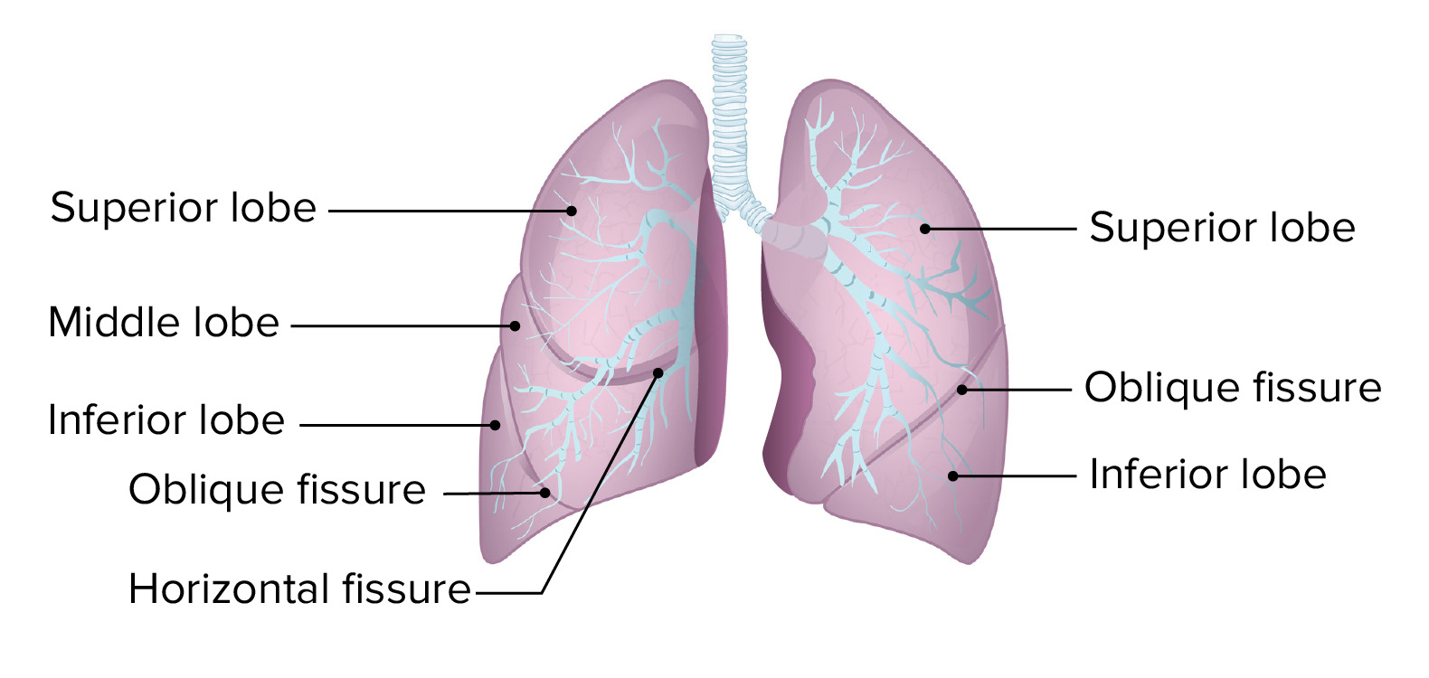
00:01 So when you first sat down to learn about the thorax, I'm guessing your first thought was, "Hey, I'm going to learn about the lungs and the heart." Iit might be a little surprise, we're just now getting to the lungs. 00:11 Probably even more surprised that we're going to do it in a single session as opposed to four or five for the heart. 00:16 That's because the real complicated stuff in the lung is occurring microscopically. 00:21 The micro anatomy, the histology is really the complicated part where the gas exchange takes place. 00:27 The gross anatomy is actually not too bad. 00:30 So we're really going to see the overall shape and direction and relations of things. 00:36 And we're going to start with the left lung, from a lateral view. 00:41 The first thing we're going to point out is the pointedness. 00:44 If we look superiorly, we see that there's sort of a vague point to the lung called the apex. 00:50 Conversely, at the bottom or the inferior portion, it's much broader. 00:55 And that part we call the base. 00:58 We have an upper lobe, that's not just more superior than the other lobe, but it's also a little more anterior. 01:05 And it has a little tiny special part called the lingula. 01:10 That we'll see later is the portion of the upper lobe that sits right over the anterior aspect of the heart. 01:18 And then the other lobe is the lower lobe. 01:20 And despite the name, it's not just somewhat lower, but it's also a little bit more posterior than the upper lobe. 01:28 And that's because the gap or fissure that separates the two, it's running diagonally. In fact, it's called the oblique fissure. 01:37 If we swing around to the right, again, from a lateral view, we can see a pointy apex superiorly and a broad base inferiorly. 01:46 We still have an upper lobe, and we still have a lower lobe. 01:51 And we still have an oblique fissure running along the front of the lower lobe. 01:57 But on the right side, we have an extra lobe. 01:59 We have the middle lobe. 02:01 And this middle lobe is separated from the upper lobe by a different fissure called the horizontal fissure because it's running less diagonally. 02:11 Now, let's take some medial views. 02:13 So we'll go back to the left lung and look from a medial perspective. 02:18 And we're going to focus on really the only thing there is to see here, which is that central depression or hilum of the lung. 02:25 In three key structures here. 02:28 The first one is the left bronchus. 02:32 And we see superior to the bronchus is the pulmonary artery. 02:38 Inferior is where we find the pulmonary veins. 02:43 In order to get a sense of the relationships of the lung to what's nearby, we've kind of put in these little grooves or impressions for structures that are resting right up against the left lung. 02:55 So, the first one we see here is pretty big. 02:58 And that's because on the left side, we have a group for the aorta. 03:03 We have the arch becoming the descending aorta here. 03:07 We also have grooves for some branches of the aorta. 03:11 Namely, we have the left subclavian artery here. 03:15 And we also have corresponding veins in this area. 03:18 For example, we have the left brachiocephalic vein. 03:22 And more centrally, we have this long tube, the esophagus. 03:27 But the biggest impression here is for the heart. 03:30 And so that's the cardiac impression. 03:32 And that's because the heart is mostly off to the left side of the thoracic cavity. 03:36 We'll swing around to the right, and look at the right lung from a medial view. 03:40 Again, focusing in on the hilum and the three key structures. 03:45 And we're going to start with the bronchus. 03:47 And this is a subtle but important difference. 03:50 Here on the right side, the pulmonary artery sits anterior to the bronchus. 03:56 And it may not seem like a big deal now. 03:58 But if you were to start learning about congenital heart diseases, for example, you really want to know the relationship of the bronchus to the artery And the way to remember that, is Right Anterior Left Superior. 04:13 And what that means is on the right side, the artery is anterior to the bronchus, and on the left side, it's superior. 04:22 So, R-A-L-S, or RALS is the acronym. 04:27 That sounds like a made up word, but it's kind of isn't because RALS, R-A-L-S is actually the sound for crackles when you auscultate lung sounds. 04:36 So it's at least thematically linked. 04:39 Regardless of the orientation of the artery to the bronchus on either side, the inferior most structures are going to be the pulmonary veins. 04:48 If we put in those grooves or impressions on the right side, because of the asymmetry going on in the mediastinum, we're going to see different structures here. 04:57 So we're gonna see more venous structures. 05:00 For example, we're going to see the group for the SVC, or the superior vena cava and the right brachiocephalic vein. 05:08 This is also the side that we're going to see the azygos vein. 05:13 And of course, we're going to have a little bit of a groove for the inferior vena cava before it reaches the heart. 05:21 We do have a little bit of an artery here, the right subclavian artery, but not a lot of arterial structures. 05:28 Down the midline, and the longest tube here is the esophagus. 05:33 There is a slight impression here for the heart. 05:36 But because most of the hearts off to the left, it's a lot shallower than it is on the right. 05:42 The trachea and bronchi, already covered in separate section. 05:45 But now that we've mentioned some of the asymmetry, namely the different number of lobes and the presence of the heart. 05:52 Hopefully, if you were to go back and look at that trachea and bronchi section again, you would understand why the asymmetry is what it is there. 06:01 So now we're going to talk about lymphatic drainage of the lungs. 06:04 Lymphatic drainage is important for a lot of reasons. 06:07 And one of the reasons it's important to lungs is because the lungs are a common sight of lung cancer. 06:13 And as we learned in the breast section, lymph nodes are an important part of staging how far a cancer has spread. 06:22 Cancers in the lung can spread via the blood or the lymphatics. 06:26 However, the lymphatics are rarely confined to certain areas, because lymphatic vessels and their associated lymph nodes have to exit the hilum. 06:36 We have a predetermined path, so we know exactly where these lymph nodes draining the lungs are going to occur. 06:45 Intrapulmonary lymph nodes that are draining intrapulmonary lymphatic vessels. 06:50 And then when we reach the outside border, where we hit the bronchus, we have the bronchopulmonary or hilar lymph nodes. 07:00 If we keep going, we're going to hit the carinal lymph nodes at the carina. 07:05 The tracheal bronchial nodes at that junction of the trachea and bronchus, all the way up to the paratracheal nodes. 07:13 And for the most part, these lymphatics are going to drain on the right side into the right lymphatic duct and on the left side into the thoracic duct. 07:22 But some of these upper ones, some of these paratracheal and tracheal bronchial lymphatics might drain into their own special duct called the bronchomediastinal lymph duct, which will actually drain into the venous system directly.
About the Lecture
The lecture Anatomy of the Lungs by Darren Salmi, MD, MS is from the course Thorax Anatomy.
Included Quiz Questions
How many lobes are present in the left lung?
- 2
- 3
- 6
- 4
- 5
Which fissure separates the upper and middle lobes of the right lung?
- Horizontal fissure
- Oblique fissure
- Vertical fissure
- Longitudinal fissure
- Lateral fissure
What is the most inferior structure within the hilum?
- Pulmonary vein
- Pulmonary artery
- Bronchus
- Bronchial vein
- Bronchial artery
What is the relationship of the pulmonary artery to the bronchus in the right lung?
- Anterior
- Superior
- Inferior
- Lateral
- Posterior
Customer reviews
5,0 of 5 stars
| 5 Stars |
|
5 |
| 4 Stars |
|
0 |
| 3 Stars |
|
0 |
| 2 Stars |
|
0 |
| 1 Star |
|
0 |


