Playlist
Show Playlist
Hide Playlist
Anatomy of the Eyeball
-
Slides Anatomy of the Eyeball.pdf
-
Reference List Anatomy.pdf
-
Download Lecture Overview
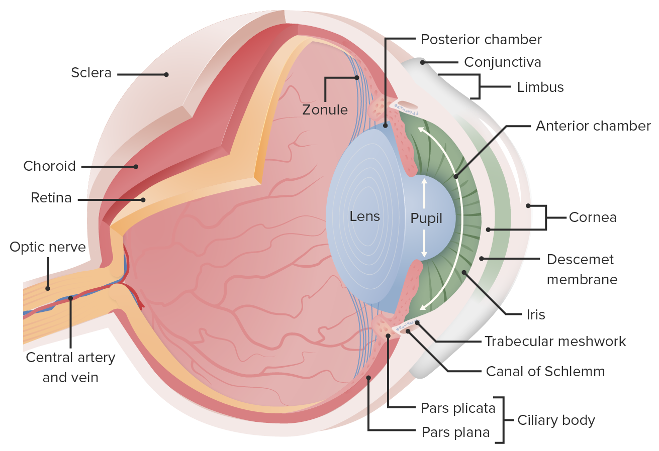
00:01 Now let's look at a sagittal cross section of the eyeball itself to see some of the small structures that lie within. 00:10 First of all, we have the fibrous tunic, which is composed of the cornea and the sclera, the cornea being very thin and the sclera being a very tough, thick connective tissue layer. 00:24 We also have the vascular tunic, which is comprised of the iris, a structure called the ciliary body at the edge of the iris and the choroid that carries much of the vascular supply. 00:38 In the middle, we have an avascular structure called the lens. 00:44 The eyeball itself can be divided into an anterior and posterior segment or cavity. 00:50 The anterior segment is smaller and contains what's called aqueous humor, and can be divided into an anterior chamber and a posterior chamber on either side of the iris. 01:04 The posterior cavity or segment is composed of something called the vitreous body, which is a much thicker gelatinous substance. 01:15 This is also where we find the retina, which is responsible for sensing visual information. 01:22 So let's take a closer look at that fibrous tunic. 01:26 So one component of that fibrous tunic was the cornea. 01:30 And the cornea is one of the few places in the body that is avascular. 01:35 And it's avascular, to help make it more transparent, so it doesn't distort any light on its way to the retina. 01:44 The other part is much tougher and thicker. 01:48 And that's the sclera. 01:50 It also doesn't have a lot of blood supply. 01:53 And that's actually what gives it its whitish appearance. 01:58 Now let's look at the vascular tunic. 02:01 We have the iris, or that circular portion that you could look at when you look at someone's eyes and it's often where we find eye color. 02:10 We have this structure called the ciliary body that it's surrounds the iris. 02:16 And then we have the vascular layer called the choroid, where much of the blood supply comes from. 02:25 So if we were to look at the eye from an anterior point of view, we would again see the iris and the space in the middle, which is the pupil. 02:35 And the iris can act on affecting the size of this pupil. 02:39 In situations where the lights are bright, the iris can constrict that pupil and it can also dilate the pupil in areas of dim light in order to let more light through and hit the retina. 02:57 Back to a sagittal view, we have the cornea, the ciliary body, and the ciliary body, and the lens. 03:06 And the lens is held in place by these suspensory ligaments. 03:11 If we're to look at this area sitting in front of the lens, we can divide the fluid here into two chambers. 03:19 We have the anterior chamber that sits anterior to the iris and the posterior chamber that sits posterior. 03:28 Now let's look at the much larger cavity the posterior cavity also called the vitreous cavity. 03:36 So everything posterior to the lens is going to be called the posterior or vitreous. 03:42 And we say vitreous because it has something called vitreous humor, which is much thicker than the aqueous humor of the anterior cavity. 03:53 And this is going to sit directly on the retina. 03:57 Therefore, any movement of this thick vitreous can have an effect on pulling in a sense the underlying retina. 04:05 So let's look at that aqueous humor and how it flows. 04:11 So here we have the ciliary body, which again surrounds the iris. 04:17 And this is where we're going to reduce the aqueous humor initially behind the iris therefore in the posterior chamber, it will flow out and overpass the pupil and reach the anterior chamber to fill it. 04:31 And then drainage for aqueous humor will be at this scleral venous sinus, also going by the eponym, the canals of schlemm. 04:41 And this is a very important area because without proper drainage of the canals of schlemm, the amount of aqueous humor could build up and create too much pressure. 04:50 And that's essentially the etiology of glaucoma. 04:57 Now let's look at the retina, the part that's actually perceiving vision for us. 05:05 Here we have the optic part of the retina, the part that's going to actually have photoreceptors and sense the changes in patterns in light. 05:14 And then there's the non visual retina that will sit beyond this border called the ora serrata. 05:22 A certain part of the retina is going to have a very high concentration of certain types of photoreceptors. 05:28 And we call that the fovea centralis. 05:30 And that's going to be the area where we sense a real clear, sharp central vision. 05:36 And then we're going to have the optic disc, which is the area where all of the information from the retina is coalescing to feed into the optic nerve. 05:48 In terms of the microstructure, if we're going from outside in, we're going to start with that fibrous tunic, the sclera, deep to which we'll have the vascular tunic, the choroid, and then the retina. 06:03 And we're going to have photoreceptors, bipolar cells, ganglion cells, and all of this will lead up to the optic nerve. 06:16 Just above the choroid layer separated from the retina is a little membrane called Brooks membrane. 06:23 And then an area of pigmented epithelium. 06:29 Then we're going to have the processes of the photoreceptor cells and photoreception is actually working outside in, in the sense that the photoreceptors are actually on the layer closest to the retinal pigmented epithelium. 06:46 Then there's a tiny membrane, called the outer limiting membrane separating it from the actual nuclei of these photoreceptor cells. 06:56 Then these areas of connection called the outer plexiform layer, giving rise to the nuclei of bipolar cells, which are therefore communicating the information from this photoreceptor cells up to the inner plexiform layer, where we have the nuclei of what are called ganglion cells. 07:17 And finally, the nerve fiber layer, which are the nerve fibers heading off along the inner limiting membrane towards the optic disc to feed the optic nerve. 07:30 And the retina has different types of photoreceptors. 07:34 They're named off of their shape, and they're the rod shaped rods, and they're sensitive to levels of illumination or how much light there is. 07:42 They're really good for night vision. 07:46 Then there are the cones. 07:48 And they're the ones used for daylight vision and specifically color vision. 07:52 Therefore, they come in three types red, blue, and green. 07:55 These are the ones that are heavily concentrated in that fovea centralis. 08:02 Let's look at the vascular supply of the eyeball itself. 08:07 Here we have the optic nerve as it enters the optic cup. 08:12 And we see that the central retinal artery and vein are traveling inside the optic nerve, hence the term central. 08:21 We also had the short ciliary arteries and the long ciliary arteries going into the choroid layer rather than the retina. 08:30 And we had drainage coming out of the vorticose, also called choroid veins. 08:38 More anteriorly, we're going to find some anastomosis. 08:43 The arteries that are supplying the extra ocular muscles will actually enter the eyeball here in anastomose at an area called the major arterial circle of the iris. 08:55 There's also a smaller minor arterial circle. 09:01 Here's a view as if you were looking at an eye during an ophthalmic exam. 09:06 What you would see in the center of your view would be this macula lutea. 09:11 And the center of which would be the fovea centralis, which is going to be a little more pigmented than the surrounding retina. 09:20 Just off to the side is where you would actually find the optic disc. 09:24 The optic disc being where all of these nerve fibers are coalescing to form the optic nerve wouldn't be an area that has photoreceptors, therefore, there's no actual vision taking place in this little area. 09:38 What you can see is all of these blood vessels are heading towards that area. 09:43 Because of the relationship of the central retinal artery and vein to the optic nerve. 09:48 So we see superior nasal arterioles and venules, inferior nasal ones, superior temporal ones, and inferior temporal ones, just based off of their location. 10:05 We also have some branches close to the macula. 10:09 And those are therefore called the superior macular arterioles and venules and the inferior macular arterioles and venules. 10:20 Here's another view as if you were looking from an ophthalmoscope. 10:25 And you can again find this macula and fovea based off of the darker pigmentation. 10:32 More nasally, we find the very bright optic disc. 10:37 And again, it's bright here because there is no retina covering it. 10:40 This is where all the nerve fibers are coalescing. 10:44 Based off the location, we can call these blood vessels, the superior nasal ones. 10:51 These ones the inferior nasal ones. 10:54 The ones more laterally as the superior temporal ones, and the inferior temporal ones. 11:00 And the ones that are closer to the macula would be the superior and inferior macular ones. 11:06 Now let's look at how light actually affects vision when it hits the retina. 11:11 As we mentioned, the cornea is a vascular in order to not distort any of the images before they actually reach the retina. 11:19 Then light will pass through the opening of the iris that we call the pupil, then through the lens, which is by convex but it is flexible and can change its shape a little bit. 11:32 And it can do that through the ciliary muscle. 11:35 And changing its shape to help things focus is a process called accommodation. 11:41 If all of that goes well, light should hit the fovea centralis, the area where we have our sharpest clearest central vision. 11:51 Then this image is actually inverted on the retina, so it's actually turned upside down initially. 12:00 Now this leads us to some problems in what we would call refraction of the bending of the light before it actually gets to the retina. 12:08 In myopia, or nearsightedness, the overall shape makes it very difficult for the image to focus where it's supposed to on the retina, making it hard to see things that are further away. 12:23 To correct this, a concave lens can actually direct the image where it needs to be. 12:31 Conversely, with what was called a hyperoptic eye or farsightedness, near vision is affected, and correcting that would use a convex lens.
About the Lecture
The lecture Anatomy of the Eyeball by Darren Salmi, MD, MS is from the course Special Senses.
Included Quiz Questions
Which of the following is part of the fibrous tunic?
- Sclera
- Iris
- Ciliary body
- Choroid
- Lens
Where is the retina present?
- Posterior segment
- Anterior segment
- Medial segment
- Lateral segment
- Anterior chamber
From where is much of the blood supply to the eye?
- Choroid
- Retina
- Iris
- Ciliary body
- Vitreous body
Where is the aqueous humor produced?
- Ciliary body
- Choroid
- Pupil
- Anterior chamber
- Scleral venous sinus
What is the innermost membrane of the retina?
- Nerve fiber layer
- Inner plexiform layer
- Nuclei of bipolar cells
- Outer plexiform layer
- Pigment epithelium
Which statement is true?
- Rods are good for night vision.
- Pigment epithelium is the innermost layer of the retina.
- Rod cells are used for daylight vision.
- Rods come in three types.
- Cones are sensitive to levels of illumination.
At which point do nerve fibers coalesce?
- Optic disc
- Macula lutea
- Fovea centralis
- Central retinal artery
- Inferior nasal arteriole
What allows for the accommodation of the lens?
- Ciliary muscle
- Cornea
- Pupil
- Anterior chamber
- Vitreous body
Customer reviews
5,0 of 5 stars
| 5 Stars |
|
5 |
| 4 Stars |
|
0 |
| 3 Stars |
|
0 |
| 2 Stars |
|
0 |
| 1 Star |
|
0 |


