Playlist
Show Playlist
Hide Playlist
Abnormalities of the Amnion, Placenta, and Umbilical Cord Part 2

-
Slides 10-64 Placenta, Fetal Membranes and Abnormalities.pdf
-
Reference List Embryology.pdf
-
Download Lecture Overview
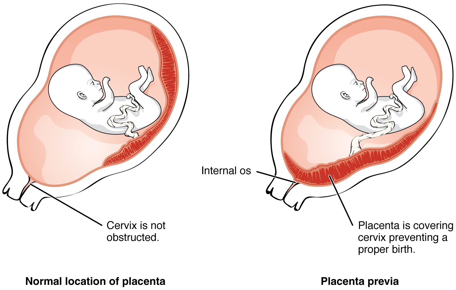
00:01 Another set of problems can occur is when we have placenta previa. 00:05 This occurs when the placenta is not where it belongs on the posterior wall of the uterus, but forms a bit lower down in the uterine cavity closer to the cervix. 00:16 It's graded in terms of severity, with the first degree being the least severe and the fourth degree being the most severe. 00:24 So in the first degree, the placenta is further down the wall than it should be, but it does not reach the internal os of the cervix. 00:32 In the second degree, it has reached the internal os but does not cover it. 00:38 No big surprise, the third degree does indeed have the placenta covering the internal os but the distinction is when the cervix dilates, the cervix is open; the placenta does not completely cover the dilated cervix. 00:53 And fourth degree, fourth degree placenta previa means that the placenta does in fact cover the internal os and it's gonna make delivery very difficult and will likely have to be done through caesarean section. 01:07 Another set of problems can arise when the umbilical cord forms in an abnormal manner. 01:13 Typically, the umbilical cord and the umbilical vessels within it enter and exit the placenta right in the middle. 01:20 When you have a marginal placenta, that simply means that the cord, instead of exiting the center of the placenta is actually somewhere around its margin. 01:28 Another issue can happen when you have a furcate placenta. 01:32 In this case, instead of the vessels converging and then leaving us the cord, the vessels leave the placenta then come together within the cord. 01:41 And last but not least is a velamentous placenta, in which case we not only have the umbilical vessels coming together outside the placenta, we have them actually attaching to the amnion and this can create additional problems during delivery and after delivery if these vessels tear and cause hemorrhage. 02:01 An issue that can arise with the development of the umbilical cord is that the vessels inside it can sometimes wrap around each other and form apparent knots. 02:10 These are actually not problematic and they're typically benign because they don't interrupt the flow of blood to and from the placenta. 02:16 However, if fetal movements cause a true knot of the umbilical cord to form, that is incredibly dangerous because the blood flow to and from the placenta and the fetus can be interrupted and result in fetal death. 02:31 So fetal movements can also cause the cord to wrap around parts of the body, this is particularly dangerous if it wraps around the neck and interrupts blood flow to and from the developing brain. 02:43 So this cord of strangulation is sometimes only noticed as a change in the movements of the fetus, so if the fetus is moving quite a bit and suddenly goes very still, it may be worth getting checked out to see if there might be some movement of the cord that's wrapped around the fetal neck. 02:59 Another problem that can occur involving the placenta occurs a little bit further back. 03:05 These are called hydatidiform moles and these are essentially a placenta without an embryo. 03:12 These form when the embryoblast inside a developing fetus, pardon me, a developing embryo, does not form and were only left with a trophoblast. 03:23 Sometimes this occurs due to polyspermy or sometimes even two spermatozoa fusing and their pronuclei attempting to create a new zygote, rather than a spermatozoa and an oocyte. 03:35 Essentially, this free forming placenta implants begin secreting human chorionic gonadotropin and mimics a pregnancy very readily, but there's no embryo present. 03:49 And during ultrasound examination, there will be an absence of the embryo noted and the villi within this hydatidiform mole will appear to be swollen and are sometimes described as looking like a bunch of grapes in appearance. 04:02 Thank you very much and we'll see you for the next talk.
About the Lecture
The lecture Abnormalities of the Amnion, Placenta, and Umbilical Cord Part 2 by Peter Ward, PhD is from the course Conception, Implantation and Fetal Development. It contains the following chapters:
- Placenta Previa
- Placenta and Umbilical Cord Anomalies
Included Quiz Questions
If the umbilical cord exits in the rim of the placenta, what is it called?
- Marginal placenta
- Furcate placenta
- Velamentous placenta
- Focal-lateral placenta
- Placenta previa
What term is used to describe hCG-secreting blastocysts that lose the embryoblast?
- Hydatidiform moles
- Trophoform moles
- Epiform moles
- Graafianoform moles
- Oophoroform moles
Author of lecture Abnormalities of the Amnion, Placenta, and Umbilical Cord Part 2

Peter Ward, PhD
Customer reviews
5,0 of 5 stars
| 5 Stars |
|
5 |
| 4 Stars |
|
0 |
| 3 Stars |
|
0 |
| 2 Stars |
|
0 |
| 1 Star |
|
0 |
