Playlist
Show Playlist
Hide Playlist
Adductor Compartment – Anatomy of the Hand
-
Slides 07 UpperLimbAnatomy Pickering.pdf
-
Download Lecture Overview
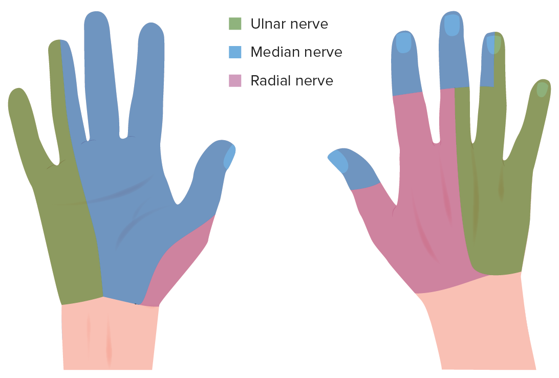
00:00 Now, let's move to the adductor compartment. 00:03 The adductor compartment only contains one muscle and this is important in adducting the thumb as the name suggests. 00:12 But there are two heads to the adductor pollicis muscle, there is an oblique head and there is a transverse head. 00:19 Here on the diagram, we can see we have this adductor pollicis muscle here, this triangular shape muscle. 00:26 It actually runs underneath here, the tendons of flexor digiti and profundus and the lumbricals which will come back to, it's running deep to those but it's passing out from the central region of the palm. 00:40 It's passing out towards the thumb and it’s got two heads. 00:44 The more transversely orientated part of the muscle is the transverse head. 00:49 The more obliquely orientated part is the oblique head and these two heads converge into a common insertion point. 00:57 The adductor pollicis, it's oblique head comes from the base of the 2nd and 3rd metacarpals and also the capitate bone. 01:06 The transverse head comes from the anterior surface of the 3rd metacarpal. 01:12 We can see the origin of the two heads. 01:15 A common insertion on to the medial side of the proximal phalanx of the thumb and this is supplied by the deep branch of the ulnar nerve. 01:25 As its name suggests, this muscles adducts the thumb, so it draws it closer towards the palm of the hand. 01:34 Now, let's move on to the hypothenar eminence. 01:39 The hypothenar eminence is located on the medial aspect of the hand. 01:45 If we can see, we've got a collection of muscles here like the thenar eminence, there are going to be three muscles within the hypothenar eminence, three muscles, abductor digiti minimi, flexor digiti minimi brevis and opponens digiti minimi. 02:05 Now, if we have a look, we can see that these muscles form a similar arrangement to that with the thenar eminence and that some are deeper to others. 02:15 Let's have a look, first of all we can see abductor digiti minimi here, and we can see where we've cut it off, it's the most superficial muscle but we can see its two parts there and then deep to this muscle, deep to abductor digit minimi, we find we have flexor digiti minimi, sometimes it's can be called brevis like I've included in the text here but as we don’t have a longus version of if, it’s not necessary and it can just be flexor digiti minimi. 02:47 And then next to it, we have opponens digiti minimi, so these are the three muscles that lie within the hypothenar eminence. 02:55 Abductor digiti minimi, you can see it's being cut here, passing along the most medial aspect. 03:01 Deep to it we have flexor digiti minimi and then slightly lateral to that, we have opponens digiti minimi. 03:09 Within the subcutaneous tissue over the hypothenar eminence, there is a small muscle, a small muscle over the hypothenar eminence and this is known as palmaris brevis. 03:23 We can’t really see it here but palmaris brevis is an an important muscle and it helps along with palmaris longus that tighten the palmar fascia and it helps supporting of the grip. 03:36 If we have to look at the origins and the insertions for these muscles, well here we have the muscles, abductor to digiti minimi, this is originating from the pisiform, bone and it inserts on to the medial side of the proximal phalanx of the 5th digit. 03:53 It's just concentrating on the 5th digit nerve. 03:56 This mostly supplied by the ulnar nerve so it's no longer the median nerve, we've moved over to the medial side of the hand and we now supply by the ulnar nerve. 04:08 Specifically, it's the deep branch of the ulnar nerve. 04:12 As the name of the muscle just suggests the function of abduct to digiti minimi is to abduct the 5th digit. 04:20 Flexor digit minimi or adding brevis if you want to originates from the hook of the hamate and also the flexor retinaculum, it shares an origin with the opponens digiti minimi muscle. 04:34 These two muscles share a common origin, however they have a different insertion, flexor digiti minimi brevis inserts again on to the medial side of the proximal phalanx of the 5th digits while as opponens digiti minimi inserts onto the medial border of the 5th metacarpal but all of these muscles have the same nerve supply which is the deep branch of the ulnar nerve. 05:01 Again, the function of these muscles is indicated by their names or flexor digiti minimi, flexes the proximal phalanx of the 5th digit and opponents digiti minimi rotates and draws anteriorly the 5th metacarpal. 05:18 It helps to oppose the little finger, and it helps to draw the little finger towards the thumb so these two digits can touch one another.
About the Lecture
The lecture Adductor Compartment – Anatomy of the Hand by James Pickering, PhD is from the course Upper Limb Anatomy [Archive].
Included Quiz Questions
The adductor pollicis muscle inserts onto which site?
- Medial side of the proximal phalanx of the thumb
- Distal phalanx of the second digit
- Lateral side of the distal phalanx of the thumb
- Lateral side of the proximal phalanx of the thumb
- Medial side of the distal phalanx of the thumb
Which muscle is present in the subcutaneous tissue over the hypothenar eminence?
- Palmaris brevis
- Abductor digiti minimi
- Flexor digiti minimi
- Opponens digiti minimi
- Flexor digitorum profundus
Which muscle originates from the pisiform bone?
- Abductor digiti minimi
- Flexor digiti minimi
- Opponens digiti minimi
- Abductor pollicis brevis
- Flexor pollicis brevis
Customer reviews
5,0 of 5 stars
| 5 Stars |
|
5 |
| 4 Stars |
|
0 |
| 3 Stars |
|
0 |
| 2 Stars |
|
0 |
| 1 Star |
|
0 |


