Playlist
Show Playlist
Hide Playlist
Accessory Sex Glands – Male Reproductive System

-
Slides 07 Human Organ Systems Meyer.pdf
-
Reference List Histology.pdf
-
Download Lecture Overview
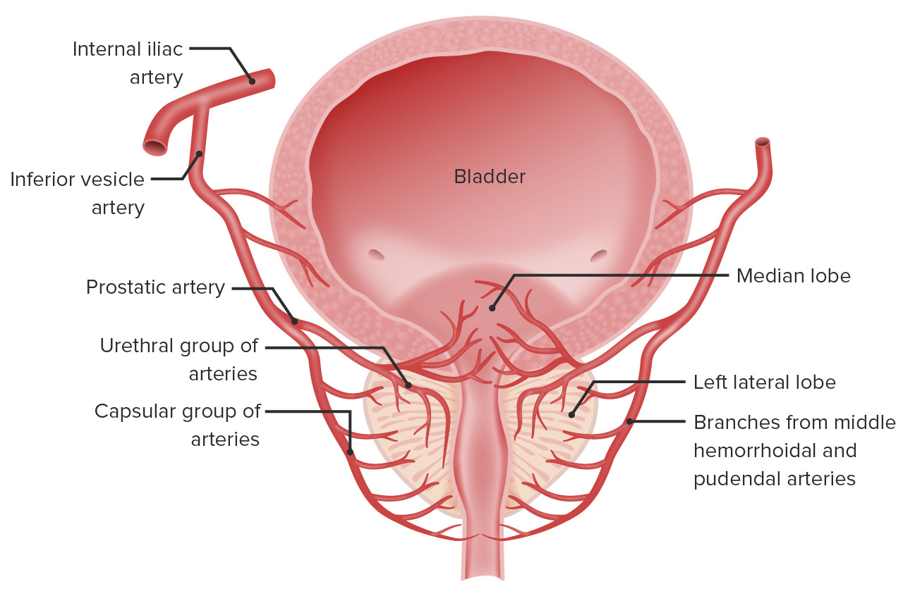
00:00 The accessory glands consist mainly of the prostate gland and the seminal vesicle, and they are the two glands that I’m going to describe now. Let’s go back to this diagram just to remind you of where the seminal vesicle lies and where the prostate lies. The seminal vesicle, you can see in this diagram, lies just at the posterior aspect of the bladder, at least from the angle you’re looking at. And the prostate gland is below the bladder. And just refresh your memories about the ejaculatory duct coming from the seminal vesicle into the prostate to join the prostatic urethra. And there, the secretion products from the seminal vesicle and the prostate will pass into the urethra during ejaculation and flash the spermatozoa out that have been delivered to the urethra by contraction of the vas deferens and also the epididymis. Let’s now have a look at this seminal vesicle in a bit more detail. Here, you can see a number of different features that are characteristic of the seminal vesicle. It has got a fairly thick muscular wall. And it has got a very, very folded mucosa. 01:31 You’ll see that more clearly on the right-hand side section taken at high magnification. 01:38 This seminal vesicle forms a very long convoluted sac, and it all connects to the ejaculatory duct. And this sac, as I mentioned, is convoluted. It’s very, very folded with lots of muscle making up the wall. The epithelium secretes fluid, and that fluid is rich in fructose which is the major energy supply for the sperm. Often, you see the seminal vesicle containing lots of colloid type material in the lumen. This reflects the fluid containing all these fructose.
About the Lecture
The lecture Accessory Sex Glands – Male Reproductive System by Geoffrey Meyer, PhD is from the course Reproductive Histology.
Included Quiz Questions
Which of the following statements about male reproductive accessory glands is INCORRECT?
- The seminal vesicles empty their secretions into the membranous part of the urethra.
- The prostate is the largest accessory gland.
- The fluid from the seminal vesicles contains fructose.
- The prostate is a dense structure that is located just inferior to the urinary bladder.
- The secretions from the prostate and the seminal vesicles pass into the urethra.
Author of lecture Accessory Sex Glands – Male Reproductive System

Geoffrey Meyer, PhD
Customer reviews
5,0 of 5 stars
| 5 Stars |
|
1 |
| 4 Stars |
|
0 |
| 3 Stars |
|
0 |
| 2 Stars |
|
0 |
| 1 Star |
|
0 |
I chose this rating because I understood the topic easily, thanks to the clear explanations and the organization of the topics
