Playlist
Show Playlist
Hide Playlist
Vulvar Intraepithelial Neoplasia & Extramammary Paget´s Disease

-
Slides Vulva Female Reproductive Pathology.pdf
-
Download Lecture Overview
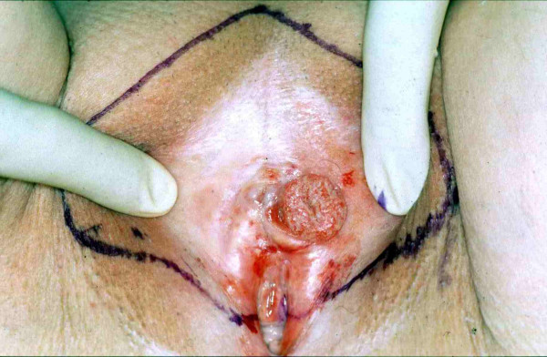
00:00 Vulvar Neoplasm Demographically, remember, because of the outside or external nature of these organs the type of cells of these organs then are outlined by will be squamous cells. 00:19 So 85% of any type of your vulvar neoplasia would have to be squamous cell. 00:26 Melanomas or adenomas could also be possible, keep that in mind. 00:30 But however, majority of a time is squamous cell. 00:33 If it is vulvar neoplasia, or think about who you are. 00:38 We still have not left the vulva, the types here would be the same strains as you do expect with your cervical cancer. 00:46 The highest strains 16, 18, 31, 33. 00:49 HPV negative associate with squamous cell hyperplasia or lichen sclerosus. 00:55 If you find you patient to be HPV-negative and the developing squamous cell cancer of the vulva. 01:03 There might be association as that I had earlier noted with lichen sclerosus. 01:14 A vulvar neoplasia that is quite different from squamous cell cancer. 01:19 Welcome to Extramammary Paget’s disease. 01:22 Put all this into perspective forming. 01:25 Mammary Paget’s disease. 01:27 Paget’s disease can be find in three different places for you on your Boards and Wards. 01:34 Paget’s disease at the bone called von Recklinghausen's disease of the bone. 01:38 Then we have Paget’s disease of the breast, mammary. 01:42 However, where are you right now? The vulva. 01:46 Therefore, you call this Extramammary Paget’s disease. 01:49 There are two major types of extramammary: the bone and the vulva. 01:54 the cell, histological will appear as being a Paget cell. 02:00 So what is Paget’s disease? What kind of cancer is this? Earlier, I had mentioned that vulvar disease if any type of cancer, 85% of time were going to squamous cell cancer. 02:14 I also said that there is a possibility of adenocarcinoma in developing. 02:19 This is an example of that. 02:21 Extramammary Paget’s disease, these are glandular cells and it’s an adenocarcinoma. 02:28 Don’t you ever forget that please? These are malignant cells confined to the epidermis as supposed to mammary or breast Paget’s. 02:36 So these are confined to the epidermis of the vulva. 02:39 Other causes of vulva neoplasia include the following: Malignant melanoma, rare but could occur in peak incidence with sixth a little bit later in age of you female, sixth or seventh decade. 02:53 Couple of important , vulvar neoplasia, make sure that you’ve understood the topic of vulvar cancer before moving on to next section. 03:02 Squamous cell cancer, HPV, higher strain, lichen sclerosus, predisposing factors perhaps. 03:08 Next, Extramammary Paget’s disease, what kind of cancer in the vulva, adeno, the malignant melanoma as where is it may be look for this perhaps in a female in her 60s or 70s. 03:21 Paget’s disease of vulva upon histologic examination will show you the following. 03:26 Clusters of large clear tumor cells within the squamous epithelium. 03:31 Clear. 03:33 These are adenocarcinoma. 03:35 Take a look at the picture on the right. 03:36 you’ll notice on the top right corner that those appears being glandular, it is an adenocarcinoma associated with pruritus. 03:48 Paget’s disease of the vulva. 03:50 Let us now take a look at what’s on the vulvar intraepithelial neoplasia. 03:54 Do not confuse it with Paget’s. 03:57 So here, with the fact that well the same type of progression that you’ll expect with cervical intraepithelial neoplasia could be found here with VIN as well. 04:07 More common. 04:08 Will progress to invasive squamous carcinoma if untreated. 04:11 Just like what you expect with CIN. 04:14 So, what is this? What would you call this? Would you call this insight too? Or would you call this dysplasia. 04:21 I’m hoping that you would tell me that this is good. 04:26 This is going to be your dysplasia. 04:29 So VIN would be increased dysplasia. 04:33 In other words, this ordered growth and in some point at time, when you go from VIN 1, 2 and 3, you will then rupture the membrane and go on to invasis squamous cancer of what organ right now? Vulva.
About the Lecture
The lecture Vulvar Intraepithelial Neoplasia & Extramammary Paget´s Disease by Carlo Raj, MD is from the course Disorders of Vulva, Vagina and Cervix.
Included Quiz Questions
Which of the following is the most common histologic type of cancer of the vulva?
- Squamous cell carcinoma
- Melanoma
- Bartholin gland adenocarcinoma
- Basal cell carcinoma
- Rhabdomyosarcoma
Which of the following statements regarding extramammary Paget disease of the vulva is TRUE?
- It involves intraepithelial glandular cells.
- It is a subtype of squamous cell carcinoma and is microscopically indistinguishable from VIN.
- It arises from squamous cells confined to the epidermis.
- It is more common than vulvar intraepithelial neoplasia (VIN).
- It is benign but can progress to squamous cell carcinoma.
Author of lecture Vulvar Intraepithelial Neoplasia & Extramammary Paget´s Disease

Carlo Raj, MD
Customer reviews
5,0 of 5 stars
| 5 Stars |
|
5 |
| 4 Stars |
|
0 |
| 3 Stars |
|
0 |
| 2 Stars |
|
0 |
| 1 Star |
|
0 |
