Playlist
Show Playlist
Hide Playlist
Upper Extremity Anatomy

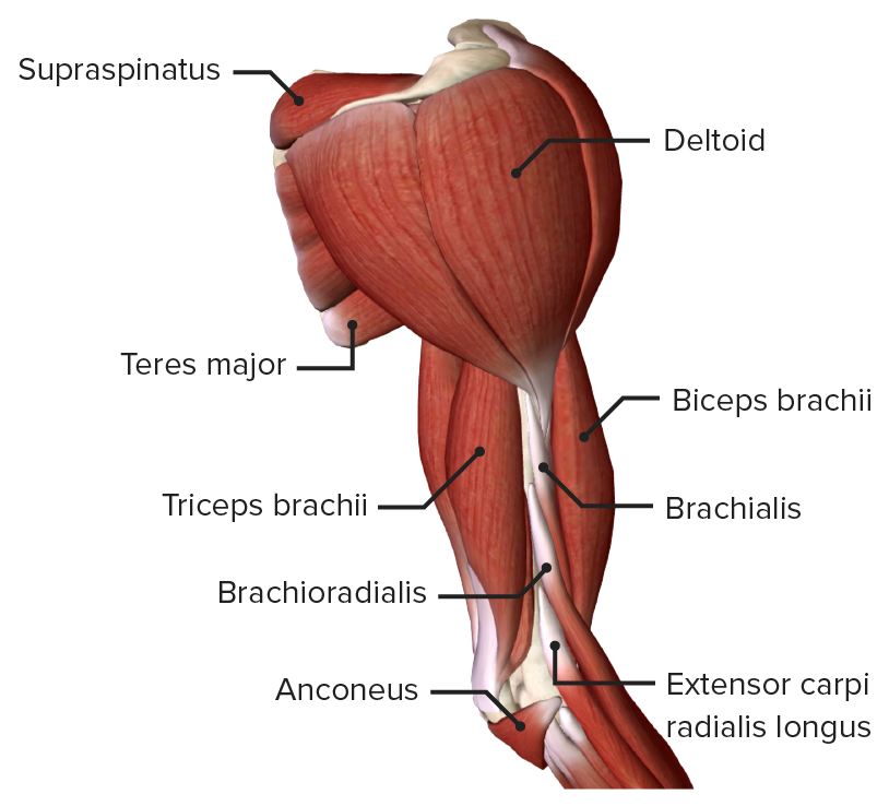
00:01 Okay, here we had the scapula humeral joint or more simply the shoulder joint. 00:07 Which parts of the skeleton articulate here? The humeral head and the glenoid fossa of the scapula. 00:14 There's only about 25% coverage of the humeral head, so dislocation is very common. 00:19 The dislocation can be posterior or anterior inferior or the head dislocates down into the axilla. 00:24 This second location is more common and the head easily travels forward under the coracoid process. 00:32 Now, where are the boundaries of the joint capsule? It extends from the glenoid to the anatomical neck of the humerus. 00:39 The anatomical neck is proximal to the greater tuberosity. 00:43 Think of a face, it's large, like our large ears on the side and the lesser tuberosity is anterior like a small nose. 00:51 The joint capsule has an axillary recess, which enables many movements in the shoulder joint. 00:56 In the anterior joint, we see the subscapularis muscle and tendon, which runs from the subscapular fossa to the lesser tuberosity. 01:04 Below this tendon there's also a subscapular recess. 01:08 Both the axillary and subscapularis recesses are important and can clearly be seen with contrast imaging. 01:13 The ligaments surrounding the shoulder are very small. 01:16 We have our glenohumeral ligaments arranged like this. 01:18 There's a superior, medial, and inferior GHL. 01:22 There's also a coracohumeral ligament which helps prevent the humeral head from subluxation downwards. 01:27 Because these ligaments are thin and weak, it's important that we have a rotator cuff. 01:33 These four muscles are the supraspinatus, infraspinatus, teres minor, and subscapularis. 01:38 All four muscles, the internal rotator and the three external rotators radiate into the joint capsule and stabilize it. 01:48 So you can think of the shoulder joint being constrained by this rotator cuff. 01:51 Of course, other muscles are also involved in the shoulder rotation, such as the deltoid, with its attachments to the clavicle, acromion, and scapular spine. 02:01 It radiates to the deltoid tuberosity, but does not enter the joint capsule. 02:05 It's more of a rotational muscle in the broader sense and not considered part of the actual rotator cuff. 02:11 Here we see the fascicles of the brachial plexus with the corresponding nerves that arise from these fascicles. 02:17 From the brachial plexus we do not see the spinal cord segments of C5 through T1, but we can see the resulting three trunks. 02:24 Each trunk divides into a ventral and dorsal portion. 02:27 The dorsal portions formed the posterior division behind the brachial artery. 02:35 And the ventral parts together form a medial and lateral divisions. 02:40 How can the student orient themselves to this? There are two ways to find the lateral division. 02:45 The first method is to search for the nerve that lies in the middle, the median nerve. 02:52 Because it's in the middle, it's formed by confluence of the medial and lateral divisions. 02:59 With this nerve identified, you can trace the median nerve proximally and laterally where you'll come to the lateral division. 03:08 The second option is to reference the coracobrachialis muscle, one of the three upper arm muscles. 03:14 It's pierced by the musculocutaneous nerve, the only nerve that originates from the lateral division. 03:21 So remember, the musculocutaneous nerve comes from the lateral cord. 03:27 Now we're here on the median nerve, which has contributions of both the medial and lateral cord. 03:32 If we go up the medial cord, we come to the medial cord branch point. 03:36 Three nerves come from the medial cord. 03:40 There's one larger nerve and two smaller nerves seen here. 03:45 The thicker one is the ulnar nerve, which travels immediately on the unlar side of the medial of a condyle of the humerus. 03:52 It lies just under the skin at this point and can be easily injured. 03:56 The two thin branches are for sensation of the upper arm and forearm. 04:00 Namely the medial brachial cutaneous nerve and the antebrachial cutaneous nerve. 04:08 To see the posterior cord we need to go behind the brachial artery. 04:12 At this point we see two nerves. 04:14 One being the axillary nerve which can be seen here. 04:16 It then pierces the lateral axillary septum here and travels backwards through the lateral axillary space. 04:24 We can explain that later. 04:27 The other nerve we find here is the radial nerve which can be seen here. 04:31 The radial nerve travels along the posterior aspect of the humerus directly on the bone between the medial and lateral heads of the tricep muscle. 04:40 This nerve can be easily injured by humerus fracture as it runs here in the spiral sulcus. 04:46 Now we look again at the posterior cord of the brachial plexus. 04:51 It lies behind the brachial artery and gives off the radial and axillary nerves. 04:57 The axillary nerve disappears here into the lateral axillary gap in the lateral axillary space. 05:04 where I'm placing the forceps. 05:07 We're now looking at it from the other side. 05:10 Here we see the forceps on the dorsal side of the shoulder blade. 05:14 We see here the infraspinatus muscle below the scapular spine, the teres minor, the teres major, and here is the long head of the triceps. 05:25 Here we have the square axillary gap and here we have the triangular medial axillary gap. 05:31 Remember, the square is lateral, triangle is medial. 05:35 A simple learning aid is to use your fingers. 05:38 You take your index and middle finger of your left hand in the index finger of your right hand. 05:42 The humerus would be the side and this would be the long head of the triceps. 05:48 Finally, this is the teres major muscle. 05:53 And up here, I have the teres minor muscle. 05:55 You can see that we have a triangular space medially in the square axillary space laterally. 06:02 In addition to the axillary nerve, the posterior humeral circumflex artery runs through the lateral axillary space. 06:09 It makes a circular arch around the posterior aspect of the humerus. 06:14 Another artery, the anterior circumflex artery runs to the medial axillary space. 06:19 Occasionally, the small subscapular nerve runs in front of the subscapularis muscle. 06:27 Here from the brachial plexus, we see the muscular cutaneous nerve, which comes from the lateral cord. 06:32 The small L for lateral looks like a one. 06:35 So remember, only one nerve comes from the lateral cord but it does also contribute to the lateral portion of the median nerve. 06:41 So we say one and a half because it's a nerve, the muscular cutaneous nerve, and here radiation namely the lateral radiation to the radial portion of the median nerve. 06:49 How does the muscular cutaneous nerve travel? Remember, it appears as the coracobrachialis muscle belly. 06:55 This can be a compression point. 06:57 And then it runs into the upper arm flexor group here and innervates the coracobrachialis, the brachialis, and the biceps brachii muscles. 07:07 It then travels distally. 07:09 And because it arises from the lateral fascicle, it supplies the lateral forum sensitivity. 07:20 Now we come to the median nerve which is in the middle of the arm. 07:24 It has a lateral contribution from the lateral cord and a medial contribution from the medial cord. 07:33 The median nerve runs along the upper arm and pierces the pronator teres muscle between the radial and ulnar head. 07:42 This is the location of pronator compression syndrome. 07:46 It then runs between the superficial and deep flexors. 07:50 Then is seen here again before finally traveling into the wrist. 08:02 It travels deep to the flex retinaculum and under the transverse carpal ligament within the carpal tunnel. 08:10 Before sending off sensory branches to the palmar surface of the radial three and a half fingers and the dorsal surface of the first three fingertips. 08:18 It innervates the forearm flexor muscles with the exception of one and a half muscles. 08:23 The flexor carpi ulnaris and the deep portion of the flexor digitorum profundus. 08:29 These one and a half muscles are innervated by the ulnar nerve. 08:32 The flexor carpi ulnaris in its entirety in the owner portion of the flexor digitorum profundus. 08:39 Both are supplied by the unlar nerve because they are in the ulnar side of the forearm. 08:45 The median nerve also supplies hand muscles primarily on the thinner aspect. 08:51 As a reminder, the median nerve supply sensitivity to three and a half fingers on the hand the thenar compartment, and the dorsal side of the distal three and a half fingertips. 09:03 It supplies motor to most of the thenar muscles and the first two lumbrical muscles. 09:09 It does not supply that the thenar muscles that pull the thumb to the ulna, the adductor. 09:16 This is a learning aid. 09:17 The muscle that pulls the thumb to the ulna is the adductor, which we can see here with its transverse and oblique heads. 09:25 It's therefore innervated by the ulnar nerve. 09:28 The muscle that pulls the thumb to the ulna is supplied by the ulnar nerve. 09:32 This is the abductor pollicis brevis. 09:34 In addition, the flexor pollicis brevis has a deep and superficial head. 09:38 The deep head lies deeper in the sense of being more ulnar. 09:41 This is why the deep head of the flexor pollicis brevis is innervated by the ulnar nerve while the superficial head is supplied by the recurrent branch of the median there. 09:49 As we discussed, it also supplies the first two lumbricals muscles. 09:53 They originate from the flexor digitorum profundus tendon. 09:57 Here we have the tendons from the flexor digitorum profundus. 10:00 Here we see the one lumbrical cut off. 10:03 and we see the second lumbrical here at the bottom. 10:06 The tendons of the flexor digitorum profundus become the first two lumbricals and are innervated by the median nerve and lumbricals three and four or by the ulnar nerve. 10:17 Now let's discuss the ulnar nerve. 10:19 It comes from the medial cord as do the medial brachial cutaneous nerve which is no longer shown here. 10:26 in the medial antebrachial cutaneous nerve, which provides sensation to the skin on the forearm and upper arm. 10:37 The ulnar nerve runs here on the medial side behind the intermuscular septum which is not shown here. 10:48 It then passes under the medial upper condyle in the groove for the ulnar nerve. 10:53 At this point, it can be easily damaged or entrapped because it lies just under the skin. 10:58 Of course it's through the medial head of the triceps. 11:01 So dislocation of the ulnar nerve can be caused by strong muscle contraction. 11:05 This is very uncomfortable and usually seen when hockey players collide. 11:08 The ulnar nerve then continues under the flexor carpi ulnaris. 11:11 This is a key muscles innervated by the ulnar nerve. 11:16 Together with the ulnar portion of the flexor digitorum profundus. 11:22 The ulnar nerve moves distally into Guyon's canal, which is not visible here between the hook of the hamate and the pisiform. 11:31 This is covered by the palmaris brevis which is also no longer visible here. 11:35 It stretches from the ulnar skin to the palmar aponeurosis. 11:38 Deep to the pisiform we have the pisohamate and the pisometacarpal ligaments which form the floor of Guyon’s canal, the ulnar tunnel. 11:50 The ulnar nerve gives off the dorsal branch of the ulnar nerve which is sensation for the ulnar two and a half fingers on the dorsal surface. 12:10 The deep branch of the ulnar nerve supplies the hypothenar muscles. 12:14 All the interosseous muscles, and the third and fourth lumbricals. 12:18 Again, that's all the interossei muscles, lumbricals three and four, and all the hypothenar muscles. 12:28 Here it gives sensation for the palmar surface of one and a half ulnar fingers. 12:36 So one and a half fingers on the ulnar palmar and then two and a half fingers on the ulnar dorsal aspect. 12:44 Here again, the dorsal branches sensation, as well as the superficial branch. 12:53 The deep branch is motor innervation for the muscles of the little fingertip, the hypothenar muscles. 13:01 The lumbricals 3 and 4, and all the interosseous muscles. 13:07 The ulnar nerve can be compressed here in Guyon's canal which is common in cyclists. 13:13 The nerve runs between the pisiform and hook of the hamate. 13:17 It's covered by a muscle that's no longer visible here the Palmaris brevis, which stretches from the ulnar skin to the palmar aponeurosis. 13:26 The floor of the pieces of form is formed by two bands, the pisohamate and the pisometacarpal ligament. 13:31 Together these borders form the Guyon's canal. 13:35 There as we said with cyclists, compression of the ulnar nerve is possible. 13:39 They experienced corresponding paresthesias and tingling in one and a half fingers on a palmar, and two and a half fingers on the dorsal. 13:50 Now, if the nerve is stimulated up here at the middle of a condyle you get a radiating paraesthesia or electrifying feeling in these fingers as well. 14:08 This is sometimes called hitting your funny bone because the patient shouts and feels the sensation of a mouse running down their arm. 14:18 Behind the brachial artery lies the posterior cord. 14:23 This gives off the axillary nerve that we see here. 14:28 As well as the radial nerve. 14:33 The axillary nerve runs through the quadrilateral space. 14:36 This is the square space remember? It's formed by the long head of the triceps medially the teres major muscle inferiorly, the teres minor muscles superiorly in the humerus laterally. 14:52 All of which together form a square. 14:56 And here we see the axillary nerve as it's intervening the deltoid and teres minor muscles. 15:04 It also supplies sensation to the deltoid skin via the lateral superior brachial cutaneous nerve. 15:10 Remember, the nerve travels around the back of the proximal humerus. 15:14 So in the case of humerus fracture at the surgical neck distal to the greater and lesser tuberosities, the axillary nerve can be damaged. 15:23 The radial nerve runs behind the brachial artery and can be seen here in the spiral groove along the back of the humerus between the medial and lateral heads of the tricep. 15:35 With midshaft humerus fractures, it can be easily injured. 15:40 It then travels distally here where we see it again. 15:43 Now, as a pullback, proximal and distal, it'll come out here on the form extensor side. 15:49 Its trunk also supplies the brachioradialis and extensor carpi radialis longus because they're still on the radial side of the humerus. 15:57 Then it splits into two branches. 16:00 The deep and superficial radial nerve. 16:04 The deep radial nerve pierces the supinator muscle here. 16:07 It can be compressed at this point in the connective tissue. 16:11 It then dives deep and innervates all the forearm extensors So the trunk supplies the brachioradialis and extensor carpi radialis longus because they originated the proximal humerus while the deep branch applies all the other forearm extensors.*** The superficial radial nerve uses the brachioradialis muscle as a guide, and supplies the sensitivity to the two and a half fingers, radio dorsally with the exception of the distal fingertips. 16:45 These are still supplied by the median nerve coming from the front. 16:49 Now we come to the six shoulder muscles. 16:52 Here we see the back of the scapula. 16:54 Above the spine on the supraspinous fossa we see the supraspinous muscle. 16:59 In here we see the infraspinous muscle below the spine. 17:02 Here the teres minor and teres major muscles as well. 17:07 Here we see the deltoid muscles laterally and anterior to the scapula originating from the subscapular fossa we see the subscapularis muscle here. 17:25 The subscapularis muscle is innervated by the subscapular nerve from the supraclavicular portion of the brachial plexus. 17:36 The muscle then runs deep to attach the lesser tubercle. 17:42 This function is internal rotation and it is in fact the strongest internal rotator of the shoulder joint. 17:49 It's also rotator cuff muscle and helps protect and stabilize the shoulder joint. 17:53 Remember the shoulder joint is maintained by weak ligaments and therefore needs muscular stability. 17:58 The inferior portion of the muscle can adduct the arm. 18:02 So internal rotation and adduction. 18:08 The supraspinatus and infraspinatus muscles are both innervated by the suprascapular nerve, which also arises from the supraclavicular part of the brachial plexus and can be seen here. 18:19 It runs under the transverse scapula ligament which bridges the scapular notch. 18:28 The supraspinatus muscle originates in the supraspinous fossa. 18:31 It runs here onto the acromion under the coracoacromial ligament in the subacromial space and insert on the upper facet of the greater tuberosity because it lies above the abduction access it abducts the arm. 18:46 And because it lies behind the axis of rotation it's an external rotator. 18:52 The infraspinatus muscle originates from the infraspinatus fossa. 18:56 It travels down towards the middle for a set of the greater tuberosity where it inserts. 18:59 It also does external rotation and its lower insertion allows it to assist with adduction because it lies below the adduction axis. 19:07 The supraspinatus above abduction. The infraspinatus below adduction. 19:15 The teres minor muscle runs from the lateral scapular border to the lower facet of the greater tuberosity. 19:21 Just like infraspinatus, it does external rotation and adduction. 19:26 As I said earlier, the teres major muscle which splits off from the latissimus dorsi is innervated by the thoracodorsal nerve. 19:34 It runs medially past the humerus and inserts on the crest of the lesser tubercle. 19:40 It therefore causes an internal rotation. 19:43 So the major does internal rotation, the minor does external rotation. 19:50 And those were the two muscles here together with the long heads of the triceps forming the quadrilateral and triangular space. 20:00 Here we see the deltoid muscle. 20:03 The deltoid muscle like the teres minor is innervated by the axillary nerve. 20:07 It originates from the scapular spine, the acromion, and the anterior clavicle. 20:18 The insertion here is on the lateral aspect of the humerus on the deltoid tuberosity. 20:23 The acromial portion performs abduction because it lies above the abduction access. 20:29 The scapular portion lies posterior causing extension, abduction, and external rotation. 20:37 The clavicular portion lies anteriorly, allowing to form internal rotation, flexion, and abduction. 20:43 The deltoid as the primary abductor of the arm above 20 degrees. 20:48 Zero through 20 is primarily supraspinatus. 20:51 Above 120 degrees, the scapular rotation and external rotation of the humerus are necessary to prevent bony impingement and allow full abduction of the humerus. 21:00 If the deltoid fails, such as with an axillary nerve lesion, then the humeral head can subluxate. 21:06 Usually the head subluxation is inferior relative to the glenoid. 21:16 Here we see the upper arm extensor. 21:18 the triceps brachii muscle. 21:21 Here is the long head with the other heads traveling to the medial and lateral sides. 21:25 Here we can see them more clearly. 21:26 This is the lateral head. 21:28 And if we look deeper, we see the medial head. 21:31 The long head is the only one that crosses two joints. 21:34 This means that it works in the elbow joint and in the shoulder joint. 21:38 The innervation of the triceps is via the radial nerve. 21:43 The radial nerve comes from the posterior cord supplies the posterior muscles of the upper arm, the triceps brachii, and then travels distally between the medial and lateral heads and the spinal groove the humerus. 21:53 Again, it can be easily damaged with a midshaft humerus fracture at this point. 21:58 The origin of the long head is the inferior glenoid on the infraglenoid tubercle. 22:11 Here you can see the fibers running down and with contraction it allows extension and adduction of the shoulder joint. 22:22 The lateral and medial heads have no effect on the shoulder joint because they originate on the humerus. 22:27 The medial head originates more distally while the lateral head is slightly more proximal and lateral. 22:32 Between them as the spiral groove. 22:35 The long, medial, and lateral heads unite to form the triceps and have a common insertion on the olecranon of the ulna. 22:43 Function therefore is extension of the elbow joint. 22:52 Here we can't see the smalliconium muscle splitting off the medial head. 22:56 It also can extend the elbow and prevents pinching of the joint capsule during extension of the elbow. 23:03 We now come to the upper arm flexors. 23:05 There are three primary muscles. 23:07 The biceps brachii, the brachialis, and the coracobrachialis. 23:11 All three are innervated by the musculocutaneous nerve from the lateral cord. 23:15 Here we see the lateral contribution of the median nerve. 23:18 Here's the lateral cord and here's the musculocutaneous nerve. 23:22 It moves into the coracobrachialis muscle, pierces it, and then travels down to innervate the short head and long head of the biceps brachii. 23:33 The short head looks longer because the long head travels through the joint capsule of the shoulder joint. 23:40 Down here we still have the brachialis muscle. 23:45 The coracobrachialis as the name suggests, has its original in the coracoid process and it travels medially inserting interior medially midway down the humerus. 23:55 In terms of its function, it's also sometimes referred to as one of the belt knotter muscles. 23:59 It does adduction, flexion, and internal rotation. 24:03 The adduction, internal rotation, and flexion. 24:09 Now the biceps brachii muscle has a short head which originates from the coracoid process. 24:15 It travels down and unites with the long head. 24:21 It has the same function as the coracobrachialis muscle in the shoulder joint. 24:24 Flexion, internal rotation, and adduction. 24:29 The long head which appears shorter here comes from inside the shoulder joint. 24:33 It originates from the supraglenoid tubercle runs through the shoulder joint capsule and passes between the greater and lesser tubercle within the intertubular sulcus. 24:42 This sulcus is is covered by the transverse humeral ligament. 24:46 It then runs distally combining with the short head and ultimately inserts onto the radial tuberosity and superficial biceps aponeurosis. 24:55 The aponeurosis is also called the lactose fibrosus and it radiates the antebrachial fascia. 25:05 The function of the long head is forearm supination, as well as shoulder flexion and abduction. 25:20 The short and long heads flex the elbow joint and supinate the forearm as the radial tuberosity is medial to the radius. 25:27 Importantly, this supination is strongest when the elbow joint is bent. 25:37 The biceps is the strongest supinator of the forearm. 25:41 Again, the biceps brachii muscle can flex the elbow joint. 25:49 Because the insertion is on the medial radius at the radial tuberosity, it can supinate. 25:55 With the elbow extended, the bicep loses the ability to supinate. 25:58 The more one bends, the better the supination effect becomes with the maximum when the elbow joint is bent at 90 degrees. 26:04 We use this advantage subconsciously when screwing in a screw with the screwdriver. 26:09 In addition, we have the aponeurosis, the lactus fibrosus which conjoined with the antebrachial fascia. 26:16 We see here the vessels are covered. 26:19 There are some cases where the lactus fibrosus is too strong and can lead to circulatory disorders as well as paraesthesiais and the distribution of the median nerve. 26:29 Here we see the brachialis. 26:33 It's originally on the medial side of the humerus opposite the insertion of the cork or brachialis. 26:43 It travels down to the ulna and serving on the ulnar tuberosity. 26:48 Its function is primarily elbow flexion, but it does help prevent the elbow joint capsule from being pinched when bending. 26:56 We now come to the three forearm extensor muscles. 27:00 First, the radial extensor group that runs down the radius laterally. 27:06 Then we have the superficial group of extensors that runs towards the ulna and little finger. 27:14 And finally, the deep forearm extensor group that runs on the radial and thumb side. 27:19 The radial extensor group consists of the brachioradialis muscle, the extensor carpi radialis longus, and the extensor carpi radialis brevis muscle. 27:28 The brachioradialis originates most laterally from the supracondylar ridge of the distal humerus. 27:33 Then slightly distal, you find the origin of the extensor carpi radialis longus and then the extensor carpi radialis brevis. 27:41 As all three run over the elbow, they assist with elbow flexion. 27:48 The brachioradialis, as the name suggests, inserts onto the radius and thus has no effect on the wrist joint. 27:55 Conversely, the extensor carpi radialis longus inserts on the base of the second metacarpal. 28:01 The extensor carpi radialis brevis inserts on the base of the third metacarpal. 28:06 By crossing the wrist and hand joints behind the flex x axis they function in wrist extension. 28:13 They also can secondarily assist in fists closing. 28:16 When the wrist is extended, the flexors are given a mechanical advantage. 28:20 This allows a much stronger finger flexion. 28:23 To remember this, you can say longus and brevis are the two fists in the radial extensor group. 28:30 In addition, the brachioradialis also performs pronation and supination because it crosses the axis of rotation of the forearm. 28:38 This axis of rotation runs from the radial head downward diagonally through the ulnar head. 28:45 The brachioradialis can supinate and if the other muscles bring the forearm into hyper supination, it can pronate the forearm back to neutral so it's capable of pronation or supination depending on the starting position of the forearm. 28:59 The extensor carpi radialis longus can also have a small supination effect. 29:03 This is sometimes called spoon supination like when one is eating soup. 29:10 The extensor carpi radialis longus pulls more to the radial side here, and it's involved in radial abduction. 29:20 One more point. 29:22 Flexion here is mainly done with a pronated forearm. 29:27 If we hang on a horizontal bar while supinated the primary muscle of elbow flexion will be the biceps. 29:39 So elbow flexion while the forearm is in pronation allows the radial forearm extensors to play a larger role. 29:47 So, supination on a horizontal bar trains primarily the biceps and the brachialis. 29:53 Now let's discuss the superficial group of forearm extensors which traveled towards the little finger on the ulnar side. 30:00 There are three muscles in this group. 30:02 All of which originate from the lateral epicondyle. 30:07 The superficial group has a more distal origin than the previous group. 30:14 This ultimately prevents them from having any significant function across the elbow joint. 30:21 The first muscle is the extensor digitorum communis which runs down here. 30:26 Approximately, it's mostly joined by the extensor indicis. 30:31 Here, between them, we often find the so called intertendinous connections. 30:36 The tendons also have different insertions. 30:38 Some insert on the second finger, sometimes even partially on the first. 30:43 Some of the tendons insert on the fourth finger, and sometimes partially on the fifth. 30:47 It can vary. 30:48 So just remember, the extensor digitorum communis with its accessory connections function as finger extension. 30:55 Some argue they do very slight extension in the elbow joint but this is nominal at best. 31:00 The next muscle is the extensor digiti minimi, which we see here. 31:06 The little finger wants to be something special, but it only has one muscle, hence extensor digiti minimi. 31:13 The third muscle is the extensor carpi ulnaris muscle. 31:16 If a muscle is called carpi, it means it inserts onto the carpus. 31:21 This muscle inserts onto the base of the fifth metacarpal. 31:24 The extensor carpi ulnaris, the extensor carpi digiti minimi, and the partial extensions of the extensor digitorum communis can in addition to extension also do ulnar deviation or moving the hand towards the ulna. 31:44 Now let's discuss the five deep forearm muscles, which run towards the thumb. 31:50 The first is named the supinator. 31:52 It acts at the elbow joint and has the strongest supination with the elbow fully extended. 31:57 Now another special consideration. 32:00 Here we see the radial nerve. 32:02 It has a superficial branch which provides sensation on the dorsal radial aspect of two and a half fingers. 32:08 The deep branch on the other hand pierces the supinator muscle through the connective tissue called the supinator arcade. 32:14 This point represents a potential impingement location for the nerve, resulting in supinator compression syndrome. 32:20 The deep radial nerve then supplies almost all the muscles of the three forearm extensor groups. 32:25 So the supinator muscle is the first muscle innervated by the deep radial nerve. 32:30 The second muscle is innervated down here. 32:32 It's the most lateral or away from the body. 32:36 So, A for Away, and A for Abductor pollicis longus. 32:40 Then comes the extensor pollicis brevis followed by the extensor pollicis longus. 32:47 So A as an Away from the body or lateral, then extensor brevis, then extensor longus. 32:55 So in total we have longus, abductor, brevis extensor longus extensor, and finally the extensor indicis muscle. 33:07 Now we come back to the supinator. 33:09 into which the deep and radial nerve courses. 33:12 Remember that supinator compression syndrome can take place at the connective tissue arcade. 33:17 The supinator originates from the ulna and the humerus and inserts onto the radius which we can't clearly see here. 33:25 Its function is as follows. 33:27 When muscles in the forearm pronate the supinator becomes wrapping away that when it contracts, it can supinate the forearm with the elbow extended. 33:37 The deep forearm extensor group has its origin not only deeper from the skin, but also further distal on the forearm at the interosseous membrane and the ulna. 33:49 We see them deeply here. 33:52 The superficial group originates on the lateral epicondyle. 33:55 Interestingly, playing tennis can cause irritation at the tendon origin resulting in lateral epicondylitis or tennis elbow. 34:03 The deep group is spared because of its distal origin. 34:10 Here we have the abductor pollicis longus. 34:13 The abductor pollicis longus travels out to the thumb. 34:17 It does some abduction and extension. 34:21 Then we have the extensor pollicis brevis next to it which does some extension. 34:26 Then comes the extensor pollicis longus accordingly. 34:29 It makes a sharp turn here and this bend can lead to what's called drummers palsy. 34:35 It can tear at this point if it's overstrained. 34:39 Last but not least the extensor indicis. 34:42 So these muscles all do extension but because they're on the radial side they also assist with radial wrist deviation The abductor does abduction of the thumb and the two extensors do extension. 34:54 We now come to the superficial group of forearm flexors. 34:58 Its origin is at the medial epicondyle. 35:04 Here there may be some tenant irritation resulting in medial epicondylitis or golfers elbow. 35:11 The following muscles are the superficial forearm flexors. 35:15 First, we have the pronator teres with the two ulnar heads. 35:20 The median nerve travels between these heads and can be irritated or compressed causing pronator compression syndrome. 35:27 Next up is the flexor carpi radialis. 35:30 The tendons of this muscle act as a landmark for radial artery palpation of the wrist. 35:35 The artery lies lateral to the tendon while the median nerve passes medially prior to entering the carpal tunnel. 35:46 The next muscle is the palmaris brevis. 35:48 It attaches the palmaris aponeurosis and tightens it upon contracting. 35:53 In the ulnar side we have the flexor carpi ulnaris muscle. 35:57 Flexor carpi radialis, flexor carpi ulnaris. 36:01 In the middle we see the palmaris longus and appear the pronator teres. 36:05 The fifth superficial flexor muscle lies under these three. 36:10 And we can see it here. 36:12 This is the flexor digitorum superficialis muscle. 36:18 We can see that its tendons are split at the end and are pierced by the tendons of the flexor digitorum profundus muscle. 36:24 The flexor digitorum superficialis is therefore sometimes called the perforatus muscle. 36:33 The superficiality is perforated by the profundus muscle. 36:39 Now remember, with the superficial forearm flexor group, some can flex the elbow, others flex only the wrist. 36:50 Some calls flexions in the MP joints and some flex the proximal and distal interphalangeal joints. 36:58 In addition, this group can do radial and ulnar deviation as well as pronation. 37:07 Now, which muscle does what? The pronator teres muscle does the pronation and its humeral portion does the elbow flexion. 37:20 The flexor carpi radialis muscle can also pronate the forearm as well. 37:24 The flexor carpi radialis muscle can also pronate the forearm as well as wrist flexion and radial deviation. 37:34 The palmaris longus partially flexes the elbow joint and tenses the palmar aponeurosis. 37:46 The flexor carpi ulnaris muscle does the ulnar deviation, and flexion of the wrist. 37:56 Below them lies the flexor digitorum superficialis. 38:00 It inserts on the central phalanx and is then pierced from below from the profundus which extends up to the distal phalanx. 38:08 Since it only inserts up to the central phalanx it cannot flex the DIP joints. 38:13 It does flex the proximal interphalangeal joints, the MP joint, and the wrist. 38:18 It can do this because it has its origin on the medial epicondyle. 38:24 When it comes to the deep forearm flexor group here we have the flexor digitorum superficialis, lying atop the flexor digitorum profundus. 38:38 We've already discussed that it pierces the superficialis and inserts onto the distal phalanx. 38:43 Because of this, it's sometimes called the perforans muscle while the superficialis is the perforatus muscle. 38:50 And because it inserts on the distal phalanx, it can flex the DIP joint. 38:55 Then fingers two through five had the flexor digitorum profundus. 39:00 The thumb also needs a flexor, and that's the flexor pollicis longus. 39:04 And we see that here. 39:05 At the very bottom we have the third muscle lying between the radius and the ulna, the pronator quadratus. 39:10 So flexor digitorum profundus. 39:12 Here's the superficialis and goes the distal phalanx then the flexor pollicis longus and deeper the pronator quadratus. 39:21 The deep forearm flexor group lies more distal in the forearm. 39:25 So it's not only deeper from the skin, but also further down the arm. 39:31 It has its origin in the interosseous membrane and along the ulna. 39:35 The flexor digitorum profundus, as we've already said pierces the superficialis and ends at the distal phalanx so naturally it flexes all the joints. 39:43 In the DIP, the PIP, the MP, and the proximal wrist. 39:54 It flexes all but the elbow joint because its origin is too distal. 40:01 Here we see the flexor pollicis longus muscle again. 40:03 It also starts at the interosseous membrane and ulna and has no effect on the elbow joint. 40:09 It then inserts on the radial side of the abduction axis allowing radial wrist deviation. 40:14 It then pulls medially from the axis of the CMC joint So that the CMC joint or saddle joint it allows thumb flexion. 40:20 It then inserts at the distal tip of the thumb allowing flexion at the phalanges. 40:24 At the very bottom we find the pronator quadratus muscle again. 40:28 It courses between the radius and ulna and as the name suggests, does forearm pronation. 40:34 The forearm flexors are innervated by the median nerve which runs up to the pronator and then between the superficial and deep flexor group. 40:45 Here's where it gives off the sensation branches for three and a half fingers on the radial palmar side and the distal dorsal skin even though that's not shown here. 41:02 The only muscle that's not supplied by the median nerve is on the ulnar side. 41:05 The flexor carpi ulnaris muscle. 41:08 It's the primary muscle supplied by the ulnar nerve. 41:13 But remember the ulnar nerve also supplies the ulnar portion of the flexor digitorum profundus. 41:20 So one and a half forearm muscles are supplied by the ulnar nerve. 41:23 The rest are from the median nerve. 41:26 We now come to typography, compression syndromes, and guide muscles of the upper extremity. 41:32 A compression syndrome of the brachial plexus is the so called Thoracic Outlet Syndrome. 41:38 Like the five fingers of a hand, it's made up of five constriction points. 41:42 We can't see the scalene neck muscles but they cause interval compression between the anterior and medial scalene muscles as the brachial plexus passes through. 41:50 Second, a cervical process such as an elongated anterior tubercle of the seventh cervical vertebrae is the second common location for a lesion. 41:59 Third, one can have a costoclavicular compression syndrome between the clavicle which is shown here at the first costal rib. 42:06 Fourth, the pectoralis minor muscle here plays a role in hyperabduction syndrome. 42:11 The arm is hyper abducted and compression takes place primarily from the pectoralis minor. 42:16 And last but not least, not visible here either is the so called long armpit arch. 42:21 A connective tissue or muscular cord which can occur between the pec major and the latissimus dorsi muscle. 42:27 These are the five points of brachial plexus compression. 42:31 In this example, we can only actually see the pectoralis minor muscle causing hyper abduction syndrome.
About the Lecture
The lecture Upper Extremity Anatomy by Prof. Dr. med. Steffen-Boris Wirth (EN) is from the course Gross Anatomy: In the Lab.
Included Quiz Questions
With respect to the glenohumeral joint, which statement is NOT true?
- The joint capsule extends from the glenoid cavity to the greater tubercle distally.
- Caudal dislocation of the humerus is the most common injury.
- The joint is primarily stabilized by muscles.
- The rotator cuff muscles radiate into the joint capsule.
- The deltoid muscle does not radiate into the joint capsule.
Which statement about the axillary space is true?
- Both the teres major and teres minor muscles form boundaries for both axillary spaces.
- The lateral axillary space is bounded by three structures.
- The humeral head is a boundary for both axillary spaces.
- The brachial artery passes through the lateral axillary space.
- A nerve that originates from the lateral cord of the brachial plexus passes through the lateral axillary space.
With respect to the brachial plexus, which statement is the LEAST accurate?
- The thickest nerve from the medial cord is the radial nerve.
- Only one infraclavicular nerve is formed exclusively by fibers of the lateral cord.
- The median nerve pierces the pronator teres muscle on the forearm.
- The median nerve runs ventrally to the brachial artery.
- The coracobrachialis muscle is innervated by the musculocutaneous nerve.
If the medial cord is severed, what deficit would be expected?
- Numbness on the skin overlying the flexor carpi ulnaris muscle
- Numbness on the skin overlying the pronator teres muscle
- Oath hand
- Failure of the thenar musculature
- Sensory loss at the fingertip of the middle finger
With respect to the median nerve, which of the following is NOT true?
- It travels dorsally to the deep forearm flexors.
- It has fibers from two brachial plexus cords.
- It passes through the carpal tunnel.
- It runs along the upper arm, medial to the biceps brachii muscle.
- It supplies 3.5 fingers on the radiopalmar side.
With respect to the ulnar nerve, which of the following is the most accurate?
- Lumbricals 1 and 2 are not innervated by the deep branch of the ulnar nerve.
- Its only muscle is the flexor digitorum profundus.
- It travels laterally to the brachial artery in the upper arm.
- The hypothenar muscles are innervated by the ulnar dorsal nerve.
- Only the interossei muscles are innervated by the ulnar nerve.
With respect to the radial nerve, which of the following is the most accurate?
- Its superficial branch supplies radiodorsal skin sensation to the hand.
- Its superficial branch pierces the supinator muscle.
- Its deep branch innervates the coracobrachialis muscle.
- Its deep branch primarily innervates the brachioradialis muscle.
- Its deep branch supplies some of the thenar muscles.
Which of the following statements is the most accurate?
- The subscapularis muscle can perform adduction.
- The subscapularis muscle is an external rotator.
- The subscapularis muscle has its origin in the dorsal scapula.
- The subscapularis muscle is innervated by the suprascapular nerve.
- The subscapularis muscle is often the cause of impingement syndrome.
With respect to the upper arm extensors, which of the following is NOT true?
- The anconeus muscle acts as a supinator in the forearm during contraction.
- All extensors of the elbow are innervated by the radial nerve.
- All heads of the triceps brachii muscle have their insertion at the olecranon.
- The anconeus muscle extends from the ulna to the lateral epicondyle of the humerus.
- The radial nerve runs between the lateral and medial heads of the triceps brachii muscle.
Which of the following statements is the most accurate?
- The bicipital aponeurosis radiates into the antebrachial fascia.
- The bicipital aponeurosis runs laterally and distally from the biceps tendon.
- The bicipital aponeurosis is crossed by the pronator teres muscle.
- The bicipital aponeurosis is crossed by the brachial artery.
- The bicipital aponeurosis is crossed by the median nerve.
Which of the following statements is NOT true?
- The brachioradialis muscle performs only flexion and pronation.
- The pronation/supination axis of the forearm runs from the proximal radial head to the distal ulnar head.
- The brachioradialis muscle does not perform any movement in the wrist.
- When the hand is pronated, flexion in the elbow joint is performed primarily by the radialis group.
- All muscles of the radialis group perform radial abduction in the wrists.
Which of the following is the most accurate?
- The extensor carpi ulnaris inserts distally to the carpal bones.
- The brachioradialis muscle originates more distally than the extensor digitorum muscle.
- The insertion of the extensor carpi radialis brevis muscle is more lateral than that of the extensor carpi radialis longus muscle.
- The origin of the superficial forearm extensors is medial to the olecranon when the elbow is extended.
- The origin of the supinator muscle is the most proximal of all forearm extensors.
Which of the following muscles does NOT flex the elbow?
- Flexor digitorum profundus
- Pronator teres
- Extensor carpi radialis longus
- Flexor carpi ulnaris
- Palmaris longus
Which muscle is most likely to originate from the palmer aspect of the ulna?
- Pronator quadratus
- Flexor pollicis longus
- Pronator teres
- Supinator
- Palmaris longus
Which of the following is NOT a brachial plexus compression location?
- Between the lesser tuberosity and the acromion
- In the scalene interval
- Between the clavicle and the first rib
- Between the coracoid process and the pectoralis minor muscle
- By an accessory cervical rib
Author of lecture Upper Extremity Anatomy

Prof. Dr. med. Steffen-Boris Wirth (EN)
Customer reviews
1,0 of 5 stars
| 5 Stars |
|
0 |
| 4 Stars |
|
0 |
| 3 Stars |
|
0 |
| 2 Stars |
|
0 |
| 1 Star |
|
1 |
It was like it was made in the 1980's!Not clear at all and failing attempts to make intresting.
