Playlist
Show Playlist
Hide Playlist
Tests for Cervical and Lumbar Radiculopathy

-
Reference List Physical Examination.pdf
-
Download Lecture Overview
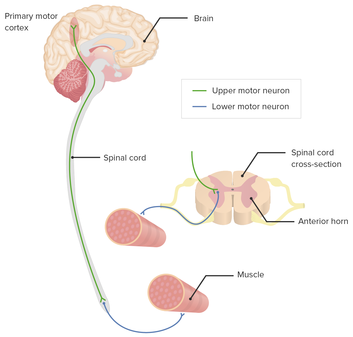
00:01 So, we've talked about some different manifestations of upper motor neuron disease, it's important for us to do a few more test that can augment our exam. 00:08 So first off, one marker of upper motor neuron disease would be a positive Hoffman sign and I'm going to demonstrate that with your right hand. 00:17 Basically what we're doing is flicking the middle finger or snapping it and you're looking for a reflex contraction in the first and second digits like so. 00:28 It can be normal if it's subtle and symmetric on both sides, but if a person is reporting paresthesias in one limb and you find the presence of a Hoffman in that limb and not in the other that would be more concerning for real upper motor neuron disease whether it's involving the cortex or something more distal to that. 00:47 That's the Hoffman maneuver. 00:48 Next stop, when we're thinking about radiculopathy in the spine involving one of the nerve roots in the cervical spine being pinched by either an osteophyte or by a disc herniation or something encroaching on the spinal canal, you can perform a foraminal compression test. 01:05 I'm going to tilt your head to the right and back a bit then I'm going to push down on your head like so, just like that. 01:16 And I applied enough pressure there that you saw him buckle a little bit, that's a sign that you've applied enough pressure. 01:22 In a person with radiculopathy, if he was having radicular symptoms going down the right arm that were coming and going, by performing that maneuver, I'd be attempting to reproduce his radiculopathy and confirm that that is in fact what's going on. 01:36 Keep in mind that I would not do that maneuver in a person who you know has rheumatoid arthritis or significant issues with spinal disease potentially a prior cervical facture, that kind of thing, better to avoid doing that kind of manipulation of the C spine. 01:54 So when we're thinking about patients who have radiculopathy, some of the classic findings you're going to find are evidence of lower motor neuron disease usually, so that would be a lower motor neuron finding. 02:04 He would have this electric shock shooting pain going down his arm because I'm pinching a radicular nerve. 02:10 He might have weakness in one of those nerve roots, he'd have hyporeflexia as well on that area and he may have sensory findings that are going down one of those dermatomes that we talked about before. 02:21 The last finding for upper motor neuron disease involving the upper extremities would be something called Lhermitte's sign. 02:26 All I'm going to do is have you tilt your chin forward and touch your chest that is something which sometimes we see in multiple sclerosis or transverse myelitis. 02:35 By him leaning forward like that he's simply tugging on his cervical spine on his cervical spinal cord, you can relax and if there is a lot of tightness or stenosis in the canal, by pulling his spinal cord over that tightness, he may develop electric shock shooting pains going all the way down his back, that would resolve as soon as he lifted his head back up again. 02:56 So having looked for findings of radiculopathy in the upper extremities, now let's looks for findings of radiculopathy in the lower extremities and we're going to start by doing our straight leg raise which is the most common maneuver that we use when folks have back pain and have any kind of sciatica type symptoms, so if you would mind just lying flat on your back. 03:21 The straight leg raise is an effort to try and put some tension on the sciatic nerve as its exiting and heading down the back of the leg. 03:29 It's passive as opposed to the patient doing it, I'm the one whose doing it, so I'm just going to make sure his leg is straight I'm going to lift it up to around 45 to 60 degrees and I'm trying to see if this reproduces any of that electric shock type shooting sensation going down to his toes, which would be consistent with sciatica. 03:46 Importantly, pain is just behind the thigh is more likely just hamstring discomfort and is not going to be indicative of a lumbosacral radiculopathy. 03:56 If somebody does have pain back there and you're not sure if it's going past the knee or not, you can actually dorsiflex the foot, that should really put tension on the lumbosacral nerves and would cause a reproduction of those electric shock shooting pains down the leg, that's called the Braggard sign or this is just called a straight leg raise. 04:14 While a passive straight leg raise on the side of the pain is useful and can be fairly sensitive, it's actually if I do the other side if I lift up the contralateral leg and that causes shooting sensations down his, down the ipsilateral lateral leg, the right side in this case, that is highly specific for having a radicular problem. 04:36 Essentially, if I may use my model, so if these are the nerve roots that are heading off to form the cauda equina and ultimately they head out and innervate various muscles in his legs, if I tug on one side and there's a lot of tightness over here on the other side where these nerve roots are exiting, simply pulling on the spinal cord and pulling it away from one side could exacerbate a foraminal stenosis on the other side. 05:03 So by lifting up his left lower extremity, if that reproduces right lower extremity symptoms, that's highly suggestive of a radiculopathy. 05:11 Lastly, for patients who have evidence of an upper motor neuron disease in the leg, back when I was checking for reflexes, if I found a four plus reflex it's good to know how to assess for clonus, so we'll demonstrate that really quick. 05:25 You're simply taking that foot that you know already had hyperreflexia and relaxing it and then I'm going to quickly pull up on the foot and you're looking for clonus which would be where his foot would have this persistent clapping down on my hand. 05:41 It's normal to have one or two beats of clonus, but if you have three, four, or five beats or even sustained clonus where there's constant flap against pressure, that would be indicative of an upper motor neuron problem concerning again for either a hemispheric stroke or potential involvement of the spinal cord. 06:01 So we were just talking about using the straight leg raise that's specifically hones in on a radiculopathy in the L5, S1 nerve roots, but if we want to assess for L2, L3, or L4 nerve roots, we use the femoral stretch test. 06:15 And to do that I'm going to have Shaun just fully extend his knee, then I am going to passively, basically fully extent his hip and I'm trying to see if this reproduces any pain, electric shock shooting pain that would be basically in the back of the thigh, potentially the lateral thigh as well. 06:35 The last test looking for upper motor neuron dysfunction is to perform the plantar reflex also known as the extensor reflex also known as the Babinski. 06:45 And all we're going to do is apply some, in a hockey stick maneuver and simply go in that kind of slow approach. 06:59 It should take a few seconds to fully navigate around the sole of the foot and you're looking for an abnormal circumstance, he would have his feet, his toes would flair upwards like this. 07:11 Whereas as you saw in his case, all his did, all he did was his toes sort of plantar flexed a bit which was completely normal.
About the Lecture
The lecture Tests for Cervical and Lumbar Radiculopathy by Stephen Holt, MD, MS is from the course Examinations of the Neck and Back Region.
Included Quiz Questions
Which is TRUE regarding Hoffman's maneuver?
- It indicates upper motor neuron involvement.
- It is performed by holding the tips of the thumb and index finger together.
- It involves flexion of the neck causing an "electric-like" sensation that runs down the back.
- It indicates lower motor neuron involvement.
- It indicates compression of a lumbar nerve root.
What is a positive Lhermitte's sign?
- It involves flexion of the neck causing an "electric-like" sensation that runs down the back.
- It indicates lower motor neuron involvement.
- It indicates compression of a lumbar nerve root.
- It is performed by holding the tips of the thumb and index finger together.
- It is seen in patients with vertebral compression fractures.
What physical exam finding is indicative of a cervical nerve root radiculopathy?
- Positive foraminal compression test
- Positive Lhermitte's sign
- Positive straight leg raise
- Positive Babinski reflex
- Biceps tendon hyperreflexia
Author of lecture Tests for Cervical and Lumbar Radiculopathy

Stephen Holt, MD, MS
Customer reviews
5,0 of 5 stars
| 5 Stars |
|
5 |
| 4 Stars |
|
0 |
| 3 Stars |
|
0 |
| 2 Stars |
|
0 |
| 1 Star |
|
0 |
