Playlist
Show Playlist
Hide Playlist
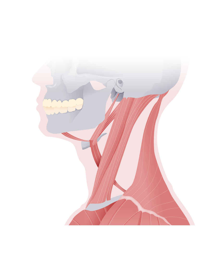
Suprahyoid Muscles

-
Slides Anatomy Suprahyoid Muscles.pdf
-
Reference List Anatomy.pdf
-
Download Lecture Overview
00:01 The next group of muscles we're going to look at are called the suprahyoid muscles. 00:06 We call them the suprahyoid muscles because they attach above the hyoid bone. 00:12 There's a similar group of muscles in the neck called the infrahyoid muscles that attach below the hyoid bone. 00:19 So here from an interior/inferior point of view, we can see the thyroid cartilage of the larynx. 00:27 The hyoid bone, and the mandible. 00:35 The first muscle we see something called the digastric muscle. 00:39 And digastric tells you a little bit about its shape because it means that we'll have two bellies. 00:47 We also see a flat muscle called the mylohyoid muscle. 00:54 An elongated one called the stylohyoid muscle and a more linear one called the geniohyoid muscle. 01:05 If we turn around to a lateral view, we can see the rest of the digastric muscle. 01:12 So here we have the anterior belly of the digastric sitting here in the digastric fossa. 01:20 And then after taking a sharp turn around the hyoid muscle goes back up towards the mastoid process via its posterior belly. 01:30 And we can see where it kind of takes this turn on the hyoid bone, there's a little thing called an intermediate tendon and that's basically what's separating the anterior belly from the posterior belly. 01:43 The innervation is somewhat interesting here in the sense that the anterior belly and posterior belly have two different innervations. 01:51 So the anterior belly is innervated by the mandibular nerve of trigeminal, whereas the posterior belly is innervated by cranial nerve VII or the facial nerve, and that just represents two different embryologic origins for these two portions of the digastric muscle. 02:10 In terms of function, it helps by opening the mouth through lowering of the mandible. 02:17 It also raises the hyoid bone. 02:20 And the posterior belly is really good at helping pull the hyoid bone upward and backward. 02:28 The next muscle we can see from this lateral view is the stylohyoid muscle. 02:35 And as the name implies, it's going to attach the base of the styloid process down to the hyoid bone, hence the name stylohyoid. 02:46 It's going to be innervated by the facial nerve or cranial nerve VII, just like the nearby posterior belly of the digastric and it is also going to pull the hyoid bone upward and backward. 03:00 Going back to more anterior/inferior view, we can see this sort of wide flat muscle called the mylohyoid. 03:09 And it's going to attach to the mylohyoid line on the medial surface of the mandible. 03:16 And of course these being the superhyoid muscles is going to attach to the body of the hyoid. 03:23 Then the fibers from opposite sides of the mylohyoid are going to meet in the midline and form a little seam or rough bay. 03:32 In terms of innervation, the mylohyoid is innervated by the mandibular branch of trigeminal or cranial nerve V3. 03:41 And it's going to help elevate the floor of the mouth as well as the hyoid bone. 03:48 Deep to mylohyoid we have the geniohyoid, and that's going to attach to the inner surface of the mandible in the area of the chin, because genio is another word referring to chin. 04:01 And of course going back towards the hyoid bone. 04:05 In terms of innervation, this is going to be innervated by a branch of the anterior ramus of the cervical one (C1) spinal nerve. 04:13 So C1 innervates the geniohyoid. 04:17 It's also going to help elevate the hyoid bone, especially when the mandible is in a fixed position. 04:24 But it can also pull the mandible downward if the hyoid bone is fixed.
About the Lecture
The lecture Suprahyoid Muscles by Darren Salmi, MD, MS is from the course Muscles of the Head.
Included Quiz Questions
What innervates the anterior belly of the digastric muscle?
- Mandibular nerve
- Facial nerve
- Oculomotor nerve
- Hypogastric nerve
- Hyoid nerve
What innervates the stylohyoid muscle?
- Facial nerve
- Mandibular nerve
- Oculomotor nerve
- Hyoid nerve
- Hypogastric nerve
What is the origin of the mylohyoid muscle?
- Mylohyoid line of the mandible
- Mylohyoid line of the maxilla
- Body of hyoid
- Mylohyoid line of the hyoid
- Laryngeal inlet
Author of lecture Suprahyoid Muscles

Darren Salmi, MD, MS
Customer reviews
5,0 of 5 stars
| 5 Stars |
|
5 |
| 4 Stars |
|
0 |
| 3 Stars |
|
0 |
| 2 Stars |
|
0 |
| 1 Star |
|
0 |
