Playlist
Show Playlist
Hide Playlist
STEMI and NSTEMI: Localizing AMI by ECG
-
Slides Cardiovascular STEMI and NSTEMI.pdf
-
Download Lecture Overview
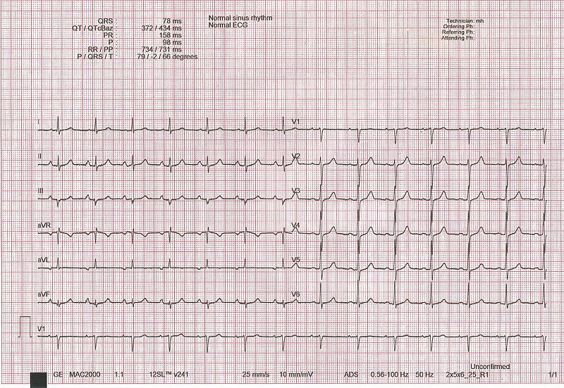
00:01 Now, localizing AMI by EKG. 00:04 If it is ST elevation, what are we going to do? We are going to localize and we will take a look at next and I have referred to this already. 00:10 Now, as I said, we are going to repeat. 00:13 In a couple of lecture series ago, I talked to you about blood vessels and their respective EKG leads, right? So now we are going to take a look at the table and we are going to harp on that very fact. 00:26 ST depression, what does that mean? It doesn't localize, but what you call an ST depression type of MI? You call that a non-STEMI, is that clear? It is not an ST elevation, it is an ST depression. 00:37 So how can you tell the difference? May I ask you this question so that you clear? How can you tell the difference between, an ST depression that you found with stable angina upon administering a stress test versus an ST depression that you might find with the myocardial infarction? You answer that question, please. 00:55 Cardiac enzymes, good. 00:57 Cardiac enzymes, are they ever found in angina? Never. Clear? Is that clear? And it should be. 01:04 Cardiac enzymes will not be found in stable, unstable angina. 01:09 Now here we go, I told you earlier that we are going to walk through the different coronary arteries and its respective ST elevations, Arteries. 01:19 I very much wish to bring to your attention that I did not say arteriole. 01:24 So let us take a look at left anterior descending, which is the artery and where exactly on the heart is the supply. 01:29 Take a look, Anteroseptal towards the apex, okay. 01:33 So, I want you to go in that order of your precordial leads. 01:37 Begin it at V1 because that is your interventricular septum, anti 2/3 of your interventricular septum is your LAD, used to be called and still is the widow maker, right. 01:47 Massive MI and you will find an ST elevation, at least, V1 through V4. 01:53 V6, I'm going to leave that out. V5, that's more lateral, but at least know V1 through V4. 01:59 Lateral walls. 02:01 Well, with lateral walls, we have left anterior descending and left circumflex. 02:07 With that said, let us take a look at ST elevation here. 02:10 If you focus on leads V1, V2, and V3 approximately in the middle of that stage of ECG, you will notice that this would be an ST elevation. 02:19 You see that crazy convex type of ST elevation. 02:24 Huge, massive, specifically in those precordial leads, which coronary artery would this be? Left anterior descending, is that clear? V1 through V4 you see something like this and please know, I don't care which licensing exam that you are taking. 02:42 All 12 leads will be given to you and when they are given to you, it is imperative that you know how to interpret and do not panic. 02:49 Once you panic, this whole thing looks like a blur. 02:51 You focus here V2, V3, V4. 02:55 What are you looking at there? An ST elevation. 02:57 And those ST elevations to you should indicate left the anterior descending type of issues. 03:04 Okay now, what if it is the lateral wall? Then it is the left circumflex also. 03:09 And this would be your V5, V6, that is official. 03:12 And we have a lead I and aVL, absolutely lateral. 03:16 Left circumflex should be your focus here. 03:18 Left anterior descending could be involved, but your focus, left circumflex. 03:22 What about the inferior wall? This would be your right coronary artery. 03:26 So, therefore, your leads here would be II at approximately 60 degrees, and from physio and from your medical education, you know lead II is probably the most important lead in terms of proper depolarization of your heart from the base towards the apex. Is it not? Of course, it is. 03:45 So lead II becomes a very important lead to you at every possible respect. 03:50 In addition to that, we have V3 and aVF, augmented foot. 03:55 And that would be the inferior portion of the heart. 03:58 And you if find an ST elevation in those leads, then you are thinking about which coronary artery? RCA. Right coronary artery. 04:07 Now here, what we are looking at is exactly that. 04:10 Take a look at lead II at the second to the bottom, you have lead II. 04:15 You have an ST elevation and for the most part, you see something like that. 04:18 Then you should be thinking, well is my patient having an inferior type of MI? And if you take a look at the aVF, you will notice there as well, you call it a fireman's hat. 04:32 If you actually take a look at that ST elevation and the QRS complex and collectively and clinically speaking, or you know just to perhaps for amusement sake, it is the fact that looks like a hat that a fireman might wear. 04:46 So ST elevations II, III, aVF. 04:50 What coronary artery, please? Right coronary artery.
About the Lecture
The lecture STEMI and NSTEMI: Localizing AMI by ECG by Carlo Raj, MD is from the course Ischemic Heart Disease: Basic Principles with Carlo Raj.
Included Quiz Questions
Complete occlusion of the left circumflex artery most likely causes ST elevation in which of the following leads?
- V5, V6, I, aVL
- V1, V2, V3, V4
- II, III, aVF, V1
- V3, V4, II, III
- V1, V2, II, aVR
What is the diagnosis in a 50-year-old man who presents to the emergency department with acute chest pain, ST segment elevation in leads II, III, and aVF, and elevated serum troponin I?
- Acute inferior wall myocardial infarction
- Unstable angina
- Infero-lateral myocardial infarction
- Anteroseptal myocardial infarction
- Right ventricular myocardial infarction
Customer reviews
5,0 of 5 stars
| 5 Stars |
|
5 |
| 4 Stars |
|
0 |
| 3 Stars |
|
0 |
| 2 Stars |
|
0 |
| 1 Star |
|
0 |

