Playlist
Show Playlist
Hide Playlist
Salivary Gland Tumors
-
Slides GIP Salivary Gland Tumors.pdf
-
Reference List Pathology.pdf
-
Download Lecture Overview
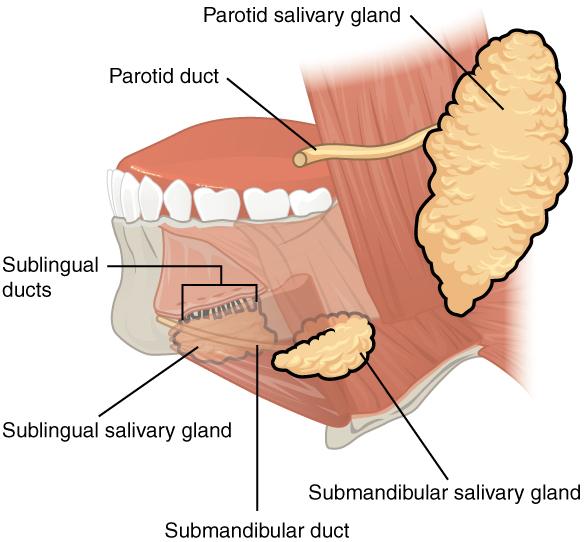
00:01 Welcome. In this talk, we're going to cover salivary gland tumors. 00:05 And fortunately, the vast majority of these are going to be benign, but there are some malignant variants. Getting to the point about benign versus malignant. 00:16 The majority, the vast majority of tumors of the salivary glands are of the benign category. 00:22 So they will fall under pleomorphic adenoma and Warthin tumors that we'll revisit in a moment and look at the way that they appear under the microscope. 00:31 There are occasional malignant tumors, so about 15% of the salivary gland tumors are going to be malignant and mucoepidermoid carcinoma is at the top of that list. 00:43 There are also things that we're not going to be talking about which regard to tumors, apparent tumors of the salivary glands and that's inflammation. 00:51 So inflammation can cause swelling, edema, some increased redness, etc., and that's not something we're going to cover in this talk. 01:01 And we're also not going to cover lymphomas which will also occur within the salivary glands but are a different entity altogether.So let's start with pleomorphic adenomas. 01:12 Again, benign and the epidemiology of this is most common of the salivary gland tumors, more common in women with a peak incidence in middle age. 01:22 It's associated with prior radiation although it doesn't have to be caused by prior radiation and very, very rarely, there can be malignant transformation. 01:32 So in all cases, you will need to excise this and send it off to pathologists to see if there is a malignant component. 01:40 The pathology, and we're not going to turn you into pathologists but just to be aware of how these things look underneath the microscope. 01:48 So of the pleomorphic adenomas, they're called pleomorphic because they're a combination of elements. 01:53 There are myoepithelial cells that surround apparent glands as you see there on the left. 01:59 There are also epithelial cells in sheets as you see on the right as well as a very loose myxomatous stroma and that can sometimes even contain cartilage or bone. 02:10 So these are the elements of a benign pleomorphic adenoma. 02:14 sorry, this is usually a slow-growing, painless, and mobile mass. 02:19 So it's not fixed to the underlying structures. 02:22 The management is surgical resection and the prognosis is very good because in the vast majority of cases, this is a benign tumor. 02:31 Let's move on to the second category of benign tumors which are called Warthin tumors. 02:35 The epidemiology of these is a little different. 02:39 So this is a second most common of the salivary tumors. 02:42 Again, it's benign. It is associated with tobacco smoking whereas the pleomorphic adenoma was not. 02:48 It's also frequently bilateral, so about 10% of the time and in an individual gland, there may be multiple foci. 02:57 It mainly involves the parotid gland as opposed to the sublingual or the submandibular glands. 03:04 The pathology is - looks a little bit different. 03:07 So on the left-hand side, we're seeing some of the imaging with a large mass involving the parotid. 03:12 But on the right hand side, we're seeing the histology. 03:16 And histology shows a palisading, that means a stacked rim of benign epithelium that surrounds a central cystic area, that's the cleared out space. 03:26 And there are frequently multiple germinal centers within it. 03:30 So it has a very characteristic look to it. It looks different than the pleomorphic adenoma which had multiple different elements. 03:36 The management, very similar except that it's involving more commonly the parotid gland and coming through the parotid gland is going to be a branch, an important branch of the facial nerve. 03:47 So for you future surgeons out there, you need to cut this out very gently to preserve that facial nerve so that people can smile and grit their teeth. 03:57 So what's shown here is actually a very interesting specimen and part of the reason that we send all these out for pathology even when we know that we - they are benign, this is a tumor, this is a mass that was in a patient's parotid. 04:09 It had both a Warthin's tumor, identified here as WT. 04:14 But it also had incidentally adjacent to it, mucoepidermoid carcinoma. 04:20 And that's what we're going to see on the next - on the next part of this talk is the malignant variants of salivary gland tumors. 04:29 So mucoepidermoid carcinoma, the malignant variant or one of the more common malignant variants of salivary gland tumors. 04:38 Epidemiology on this is that this is the most common. 04:41 There are a couple other types that we're not going to go into because they're relatively infrequent. 04:45 It's only about 15% of all salivary tumors are the mucoepidermoid carcinomas. 04:52 And again, like a Warthin's tumor, occur more frequently in the parotid. 04:58 These are interestingly enough frequently almost half of cases are associated with a chromosomal translocation that creates a fusion protein between the CRTC1 and MAML2 proteins and that affects intracellular signaling so that you get uncontrolled proliferation of the cells. 05:19 The pathology is a combination of a mucinous component, so on the left-hand side, the kind of grey pink acellular material is mucin and then, there are epithelial squamous cells and it's always a combination of those two. 05:38 On the right, we see more of the epithelial cells that have a very cleared out cytoplasm with relatively less of the mucous component. 05:47 The prognosis overall is very much dependent on the histologic grade, so whether it's very poorly differentiated or very well differentiated. 05:55 I'm not going to abuse you with that kind of information. 06:00 Don't expect that you would be able to recognize that on a slide. 06:03 That's why we have pathologists like me who will look at them down a microscope. 06:08 But depending on that degree of differentiation, the histologic grade, survival can be everywhere from 50% in five years to 90% for very well differentiated tumors. 06:18 And with that, we close with our discussion of salivary gland tumors.
About the Lecture
The lecture Salivary Gland Tumors by Richard Mitchell, MD, PhD is from the course Disorders of the Oral Cavity and Salivary Glands.
Included Quiz Questions
What is the most common malignant salivary gland tumor?
- Mucoepidermoid carcinoma
- Pleomorphic adenoma
- Warthin tumor
- Neuroendocrine tumor
- Small cell carcinoma
What describes the histology of a pleomorphic adenoma?
- A mixture of epithelial and stromal components
- A mixture of lepidic and stromal components
- A mixture of papillary and stromal components
- Mucinous and squamous mixture
- Palisading rim of benign epithelium surrounding a cystic area
What best describes the histology of a Warthin tumor?
- Palisading rim of benign epithelium surrounding a cystic area
- A mixture of epithelial and stromal components
- Mucinous and squamous mixture
- A mixture of papillary and stromal components
- A mixture of lepidic and stromal components
Customer reviews
5,0 of 5 stars
| 5 Stars |
|
5 |
| 4 Stars |
|
0 |
| 3 Stars |
|
0 |
| 2 Stars |
|
0 |
| 1 Star |
|
0 |


