Playlist
Show Playlist
Hide Playlist
Renal Clinical Anatomy: Introduction

-
Slides RenalClinicalAnatomy RenalPathology.pdf
-
Reference List Pathology.pdf
-
Download Lecture Overview
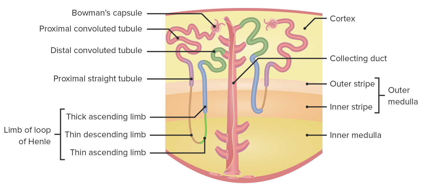
00:02 Students often have an issue with figuring out the various pathologies because they dive right into the lesions without really understanding renal clinical anatomy. So in this lecture series, initially we will be laying down the foundation as to which you need to take a look at. For example, you have heard of subepithelial, subendothelial deposits, but once again you will perhaps memorize that and at some point of time, you will be asked on electron microscopy how to identify. It is important that we lay down the foundation, in this first set of lectures so that as we move towards into glomerulonephritis and tubular interstitial disease, everything makes a lot more sense. Let us begin. We had seen this picture before and the reason we had was we were referring to secondary hypertension when dealing renal artery stenosis. Well, that was a beautiful discussion at the time. Let us now move into what we need to know here in the glomerular pathology. The glomerulus, I want you to come down the afferent arteriole and then as you do so, you are coming into the tuft of capillaries versus glomerular tuft or we will be spending a lot of time in that area and in fact, we will be blowing it up, amplifying it and then go through various imaging studies that is relevant for your diagnosis of your patient. What are the different things that are taking place in the tuft capillaries is what is relevant and our next logical question.
About the Lecture
The lecture Renal Clinical Anatomy: Introduction by Carlo Raj, MD is from the course Renal Clinical Anatomy.
Included Quiz Questions
The macula densa is an area of closely packed cells lining the wall of...
- ...the distal tubule.
- ...the afferent areteriole.
- ...the efferent arteriole.
- ...the proximal tubule.
- ...the renal capsule.
Author of lecture Renal Clinical Anatomy: Introduction

Carlo Raj, MD
Customer reviews
5,0 of 5 stars
| 5 Stars |
|
5 |
| 4 Stars |
|
0 |
| 3 Stars |
|
0 |
| 2 Stars |
|
0 |
| 1 Star |
|
0 |
