Playlist
Show Playlist
Hide Playlist
Proximal Convoluted Tubule (PCT): Correlations

-
Slides PCT Pathophysiology RenalPathology.pdf
-
Reference List Pathology.pdf
-
Download Lecture Overview
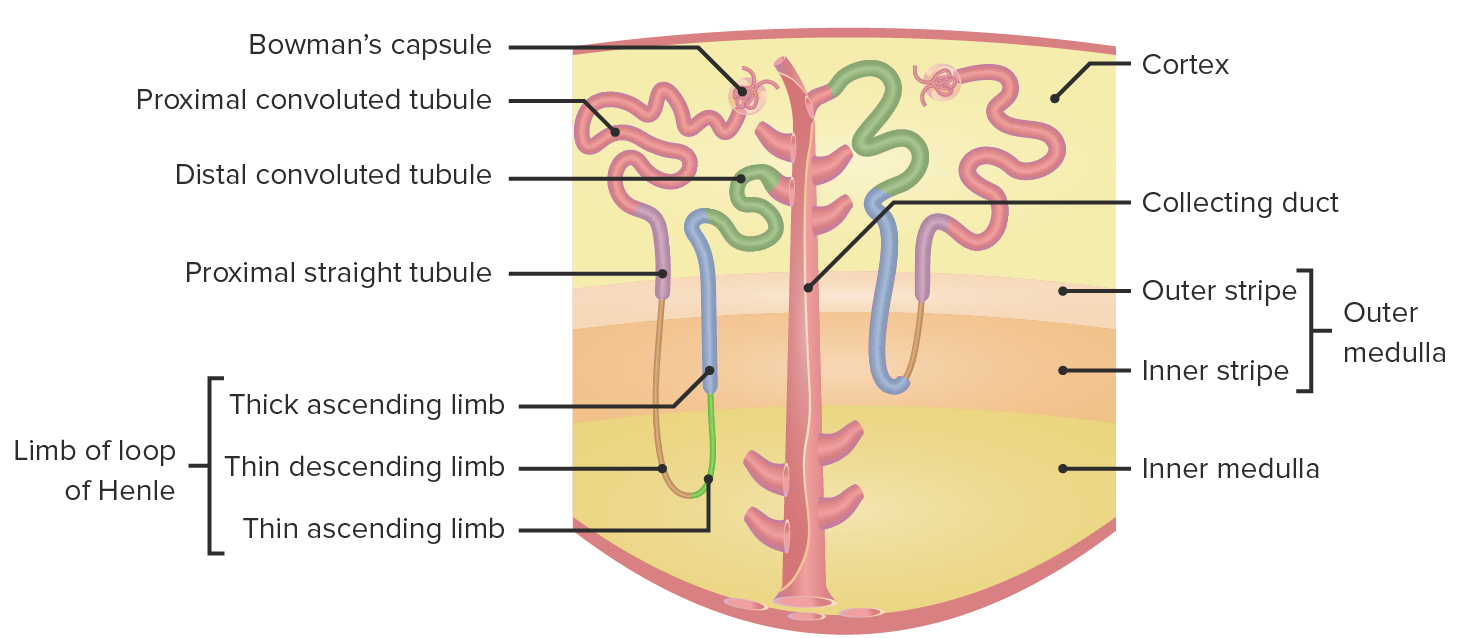
00:01 Now the PCT, the primary site of sodium reabsorption. We talked about this. Now, an important point and I actually have a separate piece of slide that will further explain upon this point. 00:14 So stop here because you can't just read through this. Okay. Bear with me. Listen, please. 00:22 Students get this confused and they keep getting this wrong on boards, what have you? Listen clinically what is it that is important for this concept? The concept here is EABV. It stands for effective arterial blood volume. Is that clear? Effective arterial blood volume. 00:45 Stop. Go back one step, foundation. What is that foundation referring to? Total body water. 00:54 Why am I bring that up? Total body water equals ECF and ICF. Of that, you tell me about ECF. 1/3 right. 2/3 is ICF. Let go ICF. I don't want that right, not relevant to our discussion. ECF yes, 1/3. What is ECF? Plasma interstitium. Tell me what is used in every single physiologic text book that you have to know now? It is the fact you have 3/4 is interstitium. What is interstitium? Tissue. What is plasma? 1/4. 01:33 That is your effective arterial blood volume. That is the most important parameter in which your baroreceptors in the carotid sinus, your juxtaglomerular apparatus in the afferent arteriole is going to be paying attention to. Correct? So let us say that you have a situation where you lose your effective arterial blood volume. How do that happen? Maybe there's hemorrhage and the blood is on the floor. You see it? Low blood. Or what else happen? The blood escaped into the interstitium. 02:16 So now effective arterial blood volume is what? Decreased, whatever reason. 02:21 If it is, now you walk me through this. If it is effective arterial blood volume that is decreased, then well the question is you decrease your GFR and you also decrease your RPF. What am I referring to when I say GFR and RPF? What formula is that? Filtration fraction. So your first instinct is if both are decreased and there must be no change. 02:42 Nope, that is not what happens clinically and that is why you are paying attention to me. So when you have a decrease in effective arterial blood volume granted, you have a decrease in GFR because a decrease hydrostatic pressure. And you have a decrease renal plasma flow because you don't have effective arterial blood volume. 02:59 So what might you want to do from the peritubular capillary? I want you to go down to the peritubular capillary now and your effective arterial blood volume has narrowed. So you have very little blood, which is coming through, into the peritubular capillary. You see the corner there. Turn the corner. Okay. So now you tell me about what is known as your oncotic and hydrostatic pressure? So what ends up happening here is that your oncotic pressure is going to be greater than your hydrostatic pressure when you have decreased the effective arterial blood volume? Why? Because you don't have as much plasma or fluid, but then you have a lot of protein. So, therefore, your oncotic pressure is quite high in the peritubular capillary. Take a look at this green line here or the green highlighted area, and you notice the P is referring to the fact that this is your plasma and your oncotic pressure is going to be much higher than the hydrostatic pressure. Where? In the peritubular capillary. So therefore, what you must keep in mind and what you must accept is that your filtration fraction is increased when you have a decrease in effective arterial blood volume and that only makes sense so that you allow for more reabsorption to take place because you have decreased effective arterial blood volume. 04:15 Now that is one example and if that physio was weak there, we are going to keep adding to it and give you specific pathologies that I have been doing here a little bit. Now let us do the opposite, shall we? Let us take a look at that decreased EABV in greater detail. It is an important concept. 04:34 Students get this confused because they either overlook it or think they have it, but they really don't. Please pay attention. So you have a decrease in EABV, why? I'll give you examples in a second. A couple of examples that I already gave you was congestive heart failure or perhaps you lost your blood, and there it is on the ground. Now all comedy aside, let us take a look at what happens seriously. You have a decrease in EABV. Okay great. And more that you have decreased in EABV, then tell me as to how was the kidney going to interpret this? Decrease renal perfusion, isn't it? So won't you think that you might want you to increase the reabsorption of your substances? That is just common sense. So as you travel through here, you have an afferent arteriole with decreased effective arterial blood volume, in other words your plasma. Don't worry about the green circle right now, I will come to that in a second. I want you to skip over to the efferent arteriole and how much do you have of your EABV? Right now it is a little bit decreased. I want you to go down to the peritubular capillary, please. If you don't have as much effective arterial blood volume, would you please tell me as to what the hydrostatic pressure is in the peritubular capillary? It is decreased, isn't it? Why? Because there is decreased effective arterial blood volume. Now if that is decreased, now the albumin or the protein never got filtered so, therefore, the oncotic pressure in the peritubular capillary, the Po that you are seeing here and that's what you are paying attention to, is then referring to the reabsorption. If you take a look at that arrow from the PCT for reabsorption, what does that mean? It means that coming out of your urine, it is extremely black and dark and bolded. And that to you should indicate there is an increase in filtration fraction when you have a decrease in EABV even though technically of a decrease in both GFR and RPF. That must be understood and I don't care how you want to do this, but at this point, I have given you an understanding as to why you have an increase in filtration fraction so that you can have an increase in reabsorption when you have a decrease in effective arterial blood volume. Now let me give you some examples clinically so that you get this. Now this part you are probably good in terms of the pathologies, but are you able to then bridge over to the physiology so that you get your questions righ. And this is where it becomes important clinically and this is where students are able to follow a question or attending to a certain extent, but then they trip over themselves because they are just not thinking enough. So examples of decreased EABV, here is congestive heart failure, cirrhosis, and hypovolemia. 07:13 I already gave you two of these examples already for decreased effective arterial blood volume. 07:19 If it is congestive heart failure, who is not functioning properly? The heart. So, therefore, what happens? Things are going to back up. What things? The plasma. Specifically, you will have your protein poor substances, which are then leaking out of your plasma into the interstitium, correct? Who is your patient? Pulmonary edema, left sided, right-sided, positive JVD and pitting edema. So there is my confirmation that fluid is leaving my blood vessel vasculature. 07:49 If you have fluid leaving the vasculature going into the interstitium, what then happens to your effective arterial blood volume? Obviously decreased. Are we clear? What happens to filtration fraction? It is increased. Reabsorption is increased in this particular setting. Cirrhosis, tell me about cirrhosis and why is it even here. Tell me as to what you look like in cirrhosis. You see my tummy. I am joking. I don't have cirrhosis. Why am I pointing to this? If there's cirrhosis, the liver is dead. Next, what happens? Let me go through the one in which albumin synthesis is not being produced adequately. So decreased synthesis of the albumin, what happens to your oncotic pressure? It decreases. Thus, what happens to your fluid? Escapes into your interstitium. What then happens to your effective arterial blood volume? Example number 2 of decreased EABV and number 3 you literally have got into state of hypovolemia, maybe there is excessive sweating or maybe perhaps there is a massive hemorrhage. What happens to effective arterial blood volume? It decreased. What happen to filtration fraction? Increased. 08:53 Do not forget that. If that is our first example of an alteration of EABV, let us take a look at the opposite end of the spectrum. This is going to be increased effective arterial blood volume, what does this mean? I want you to conceptualize this first and we will approach this exactly as to how we did with the decreased EABV. With increased EABV, effective arterial blood volume, there is more fluid passing to efferent arteriole, more across the GFR, more through efferent arteriole, more where? Through peritubular capillary. Would you please tell me about the hydrostatic pressure in the peritubular capillary? What can you predict? An increase. Take a look. Increased effective arterial blood volume. More fluid passing through. Want you to skip over your glomerulus right now. 09:50 Go to the efferent arteriole. Continue through the peritubular capillary. You told me or you followed me with knowing that your hydrostatic pressure H, you see the H, hydrostatic pressure, peritubular capillary, it is increased. You tell me about reabsorption right now. Reabsorption increase or decrease with increased effective arterial blood volume. Decreased reabsorption. 10:14 Decreased reabsorption, why? The hydrostatic pressure in the peritubular capillary is pushing. 10:20 In other words, it is offering resistance to reabsorption. So what then happens to your filtration fraction with increased effective blood volume? You are not reabsorbing. Your filtration fraction is decreased. What does that mean? Well, tell me about your blood pressure with increased effective arterial blood volume. Obviously increased. What might you want to do with your sodium? Take a look at that bottom part right there, as you pass through the PCT. See the bottom line there, it says lose sodium and water. Isn't that what you might want to do physiologically, so that you try to decrease your blood pressure? Yes, you do. So you decrease your filtration fraction. You decrease your reabsorption in the hopes of getting rid of your sodium and water so that you can perhaps decrease the blood pressure. 11:05 If you have ever been confused before, I am trying to bring your clarity. If you have never even seen this before, please make sure that you know this well and those of you that are reviewing with me and all you needed was a little bit of a trigger of things that you know, you have in the back of your head. Here it is. Make sure you know this well. Now let us take a look at examples of increased effective arterial blood volume. Say that you have mineralocorticoid excess. Give me some examples where you might have mineralocorticoid excess? You have heard of Conn's syndrome? Many times we have talked about that. You have heard of licorice? Dr. Raj what the heck does candy and a tumor in the adrenal cortex have in common? It is the fact that licorice has a component in it. Believe it or not, especially black licorice has a component in it. If you suck on a licorice that is black all of the time, you are actually be introducing mineralocorticoid into your body. Believe it or not. Believe it. Whereas if you have Conn's syndrome, that is a tumor adrenal cortex that is releasing too much aldosterone. Is there mineralocorticoid excess? Yes. I want you to go in collecting duct, what are you doing? Reabsorbing sodium and you are reabsorbing more and more water. What then happens to your effective arterial blood volume? It is increased. What happens to your hydrostatic pressure? Listen to my question. What happens to the hydrostatic pressure in the peritubular capillary? It is increasing hydrostatic pressure in the peritubular capillary. What is it doing to reabsorption? It is offering resistance. So, therefore, what then happens to filtration fraction? It is decreased because what you are trying to do? You are trying to get rid of the sodium and water. Trying to. Is that always going to be successful? It depends, right. 12:50 And then what is the other one? Isotonic gain in fluid. What happen here? Maybe this was a patient that came in who was hypovolemic. Okay. Now if you have a patient that is sweating and has been sweating severely in the hot hot sun, and you are going to bring them in. What are you going to give them immediately? IV fluids, good. What is that IV fluid that you are going to give? Right off the bat, 0,9% saline. What does that mean? 0,9% percent represents your normal isotonic saline. What if you give too much? What happens to the effective arterial blood volume? It increases. What happens to hydrostatic pressure in the peritubular capillary? It increases. What happens to filtration fraction? It is going to be decreased. Are we clear? Spend a little bit of time here, please. Make sure that you get these concepts down before you move on. It will show up. I can guarantee. 13:44 In some way, shape or form. 13:48 Here we have further, a little bit more in our discussion of the PCT with the bicarb. Luckily we have walked through this enough or we can quickly take a look at the verbiage. Okay. So here is my bicarb. Does it get filtered? Simple questions that I am asking you. Answer them. Bicarb, does it get filtered? Yes. Does it get secreted? Not at all, but majority of your bicarb in your PCT synthesized or reabsorbed? Reabsorbed. 80% of it, how? Indirectly. What do you mean indirectly? Remember this. We talked about bicarb. As soon you see bicarb, what formula is coming to mind? Carbonic anhydrase formula. Verbiage, mechanism requires bicarb joins with hydrogen and you need the help of your carbonic anhydrase. Number 1, you bring in your water and carbon dioxide into epithelial cell and with the help of your second carbonic anhydrase, you form carbonic acid and let me ask you something. For every bicarb, take a look at the bicarb in your epithelial cell inside that green box. Where is that bicarb going? It is going into the blood because you are reabsorbing it. Are we clear? My question is this. For every bicarb that you reabsorb, what are you doing to your hydrogen? You're secreting it. Are we clear? The same picture and formula can be used up in the stomach in the parietal cell that we'll do and we have done in gastroenterology. The same concept can be done in the RBC when you are dealing with the transportation of carbon dioxide. Okay. Are we good? Now with the proximal convoluted tubule, here is the exchange that you are looking for and carbonic acid then disassociates into hydrogen bicarb. The bicarb reabsorbed into the blood. For every bicarb that you reabsorbed into the blood, what are you throwing out into the urine? You are throwing out the hydrogen. What do you require for all this to occur properly? You need to make sure that you have a sodium-potassium ATPase pump on the basolateral membrane at all times to always maintain proper gradient. Let me ask you one little thing, that hydrogen those are being put out into the urine, was it a symport or antiport? Antiport, mean to say that hydrogen was doing what? It was being thrown out into the urine. 16:09 Take a look at the previous discussion, and sodium was being reabsorbed, just like you would expect. On where? The apical membrane. PCT is the place that you definitely want to pay attention because there are a lot of things that are going and that affect the entire body in numerous numerous methods as we have now outlined.
About the Lecture
The lecture Proximal Convoluted Tubule (PCT): Correlations by Carlo Raj, MD is from the course Diseases of the Nephron.
Included Quiz Questions
Which of the following fluid compartments makes up the effective arterial blood volume?
- Plasma
- Extracellular fluid
- Interstitial fluid
- Total body water
- Intracellular fluid
Which of the following conditions would not result in a decrease in the effective arterial blood volume?
- Conn’s syndrome
- Cirrhosis
- Dehydration
- Hypovolemia
- Congestive heart failure
Which of the following changes would you expect to see when there is a decrease in the effective arterial blood volume?
- Increased filtration fraction
- Increased glomerular filtration rate
- Increased renal plasma flow
- A decrease in the oncotic pressure in the peritubular capillaries.
- An increase in the hydrostatic pressure in the peritubular capillaries.
Which of the following changes is not seen in a patient with Conn’s syndrome?
- Increase in the filtration fraction.
- Increase in the hydrostatic pressure in the peritubular capillaries.
- Reabsorption of sodium.
- Decrease in the filtration fraction.
- Reabsorption of water.
Which of the following is correct regarding the proximal convoluted tubule?
- Primary site for reclamation of bicarbonate
- Primary site for reclamation of hydrogen ion
- Carbonic acetylase in involved in the formation of bicarbonate.
- For every bicarbonate that is reabsorbed, a hydrogen ion is reabsorbed.
- 80% of bicarbonate is absorbed directly.
Which of the following statements is not true regarding the metabolism of bicarbonate in the nephron?
- Bicarbonate is reabsorbed when bound to sodium.
- 80% of bicarbonate is reabsorbed in the proximal convoluted tubule.
- Bicarbonate is produced de novo in the distal convoluted tubule.
- Reabsorption in the proximal convoluted tubule occurs indirectly.
- For every bicarbonate that is reabsorbed, a hydrogen ion is secreted.
Author of lecture Proximal Convoluted Tubule (PCT): Correlations

Carlo Raj, MD
Customer reviews
5,0 of 5 stars
| 5 Stars |
|
5 |
| 4 Stars |
|
0 |
| 3 Stars |
|
0 |
| 2 Stars |
|
0 |
| 1 Star |
|
0 |
