Playlist
Show Playlist
Hide Playlist
Posterior Compartment – Anatomy of the Arm

-
Slides 05 UpperLimbAnatomy Pickering.pdf
-
Download Lecture Overview
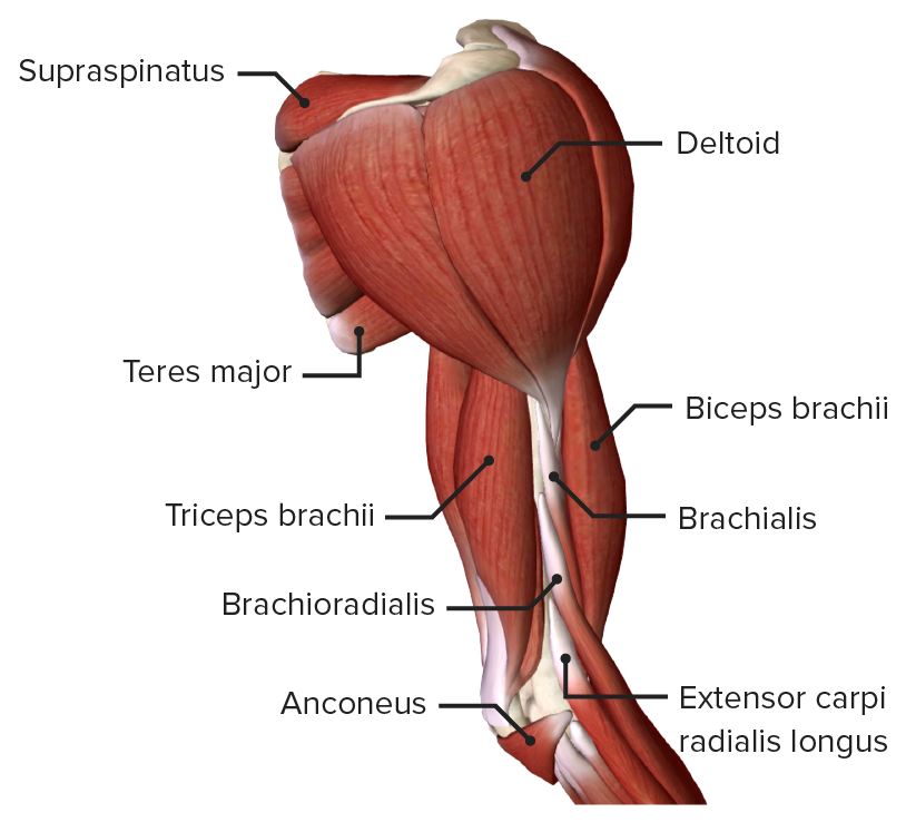
00:01 that they don't pass the needle too far and start rupturing these structures. Now let's move on to the posterior compartment of the arm. These muscles that lie behind the humerus and the median lateral intermuscular septae. What we have is triceps brachii. This muscles has got three heads, a long head, a lateral head and a medial head which we've seen some of before. 00:23 We have mentioned the long head when we covered various spaces. We can see the long head here is passing from the infraglenoid tubercle of the glenoid cavity. A long head of biceps went to the supraglenoid tubercle, while the long head of triceps is coming from the infraglenoid tubercle. It then unites to form this common tendon where the medial head and lateral head converge and all three attached on the olecranon. We've also got a small muscle here which is known as anconeus coming from the lateral epicondyle of the humerus towards the olecranon here, anconeus. But I won't really mention that. So if we look at the detail we have got triceps brachii, we have got the long head coming from the infraglenoid tubercle. We have got a lateral head and medial head. The lateral head is coming from posterior, the humerus is coming from superior to the radial groove. So if we look here, the lateral head, this is the radial groove running down here. Superior to the radial groove we have the lateral head. 01:31 Inferior to the radial groove we have the medial head. So here we can see superior to the radial groove the lateral head, inferior to the radial groove the medial head, separated by the radial groove here. We will later on see the important blood vessel, and nerves run along here. All of these three muscles converge onto the olecranon. They are supplied by the radial nerve and they are important in extending the elbow joint. So there the antagonist of brachialis and biceps. The long head because it crosses the glenohumeral joint also serves to extend the shoulder joint. I said I have mention it just briefly about we've got anconeus running from the lateral epicondyle of humerus to olecranon also supplied by the radial nerve and this really supports triceps in extending the elbow joint. 02:28 So here we've got two dissections. Two cartoons showing dissections of the posterior aspect. 02:33 Here we have got the slightly more lateral of view. We have got deltoid still intact and we can see coming from this bottom inferior border of deltoid we have got the various heads long head, lateral head and the medial head down here of triceps running towards the olecranon. 02:53 When here when deltoid has been exposed we moved deltoid, we can say that these muscles are running down towards the olecranon. We can see the olecranon here. We can see the radial groove here. We can see we have got the lateral head of triceps here and also here. What we have done is we have cut through the lateral head of triceps to expose this medial head coming from inferior to the radial groove. We have still got the long head up here. We can see it forming the quadrangular space here. We have got the long head. We have got the lateral head which has been cut is the other side of the lateral head and we have got the medial head. These all converging with anconeus on to the olecranon. So the proximal attachments of the triceps are shielded by deltoid. But removal of deltoid can highlight their attachments as we can see in this image down at the bottom. We can see the lateral intermuscular septum. 04:03 The lateral intermuscular septum here separating the anterior from the posterior compartments. 04:08 And we can also in this diagram here remind ourselves of those three spaces on the posterior wall of the axilla. So the quadrangular space we can see here, the triangular space we can see here and the triangular slit or the triangular interval we can see here. 04:25 We can also in relation to the radial groove see the origin of the medial and lateral heads of triceps.
About the Lecture
The lecture Posterior Compartment – Anatomy of the Arm by James Pickering, PhD is from the course Upper Limb Anatomy [Archive].
Included Quiz Questions
Which nerve innervates the muscles of the posterior compartment of the arm?
- Radial
- Median
- Axillary
- Musculocutaneous
- Ulnar
Which movement is performed by the muscles in the posterior compartment of the arm?
- Extension
- Flexion
- Adduction
- Pronation
- Supination
Author of lecture Posterior Compartment – Anatomy of the Arm

James Pickering, PhD
Customer reviews
5,0 of 5 stars
| 5 Stars |
|
1 |
| 4 Stars |
|
0 |
| 3 Stars |
|
0 |
| 2 Stars |
|
0 |
| 1 Star |
|
0 |
great work, u made antomy so easy especialy with the diagrams.but SIR u should discuss some clinical a bit with joints and osteology on a pre-clinical level
