Playlist
Show Playlist
Hide Playlist
Plantar Muscles of the Foot: Layer 2

-
Slide Plantar Muscles Foot Layer2.pdf
-
Reference List Anatomy.pdf
-
Download Lecture Overview
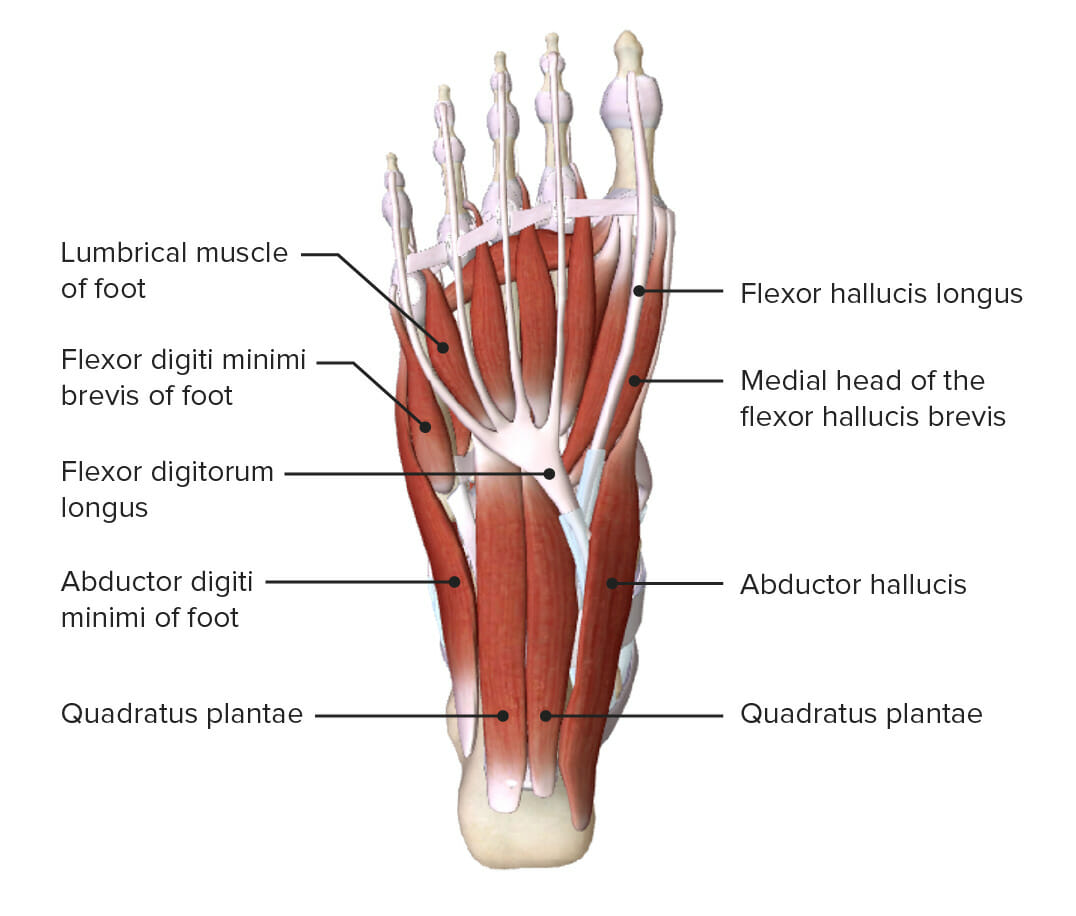
00:01 Now let's turn our attention to the second layer of muscles on the sole of the foot. 00:07 There's two muscles here, the first one is quadratus plantea. 00:10 And the second one more distally are the lumbricals. 00:14 Let's look at quadratus plantea. 00:16 The origin of quadratus plantea is the calcaneal tuberosity and the medial surface of the calcaneus bone. 00:23 It then extends distally to insert into the lateral side of the tendon or flexor digitorum longus. 00:31 Remember, flexor digitorum longus is an extrinsic muscle, so it's entered by the medial surface onto the sole of the foot and this muscle inserts into that tendon. 00:41 It's an important muscle which will serve to when it contracts to straighten and align those tendons which go on to insert onto the toe via flexor digitorum longus. 00:51 So make sure the flexion of those tendons is most efficient and occurs in a linear fashion. 00:58 So quadratus plantae is important. 01:00 Here we can see it's assisting flexor digitorum longus tendon in flexing those toes two to five. 01:08 If we then look at the lumbricals, the second muscle within this second layer. 01:13 We can see the lumbricals have an origin which is coming away from tenderness structures. 01:18 So the second third and fourth lumbricals, these come from the adjacent surface of flexor digitorum longus tendons. 01:27 So we can see these originating from those tendons or flexor digitorum longus. 01:32 The first lumbrical is coming from the medial side of that flexor digitorum longus tendon. 01:38 So the tendon is going to a specific digit. 01:41 Here, we can see the lumbricals are coming from that same named tendon but passing from that tendons surface, they pass all the way distally to the medial aspect of digits 2-5. 01:53 So these lumbricals are important in coming up the tendons and inserting onto the digits. 02:00 These muscles are involved in flexion of the metatarsophalangeal joints and also of extension of the interphalangeal joints. 02:08 So they take the same function as the lumbricals in the hand. 02:12 They obviously flex the metatarsophalangeal joints, therefore, but extend the inter phalangeal joints. 02:18 And that's just because of the position of these muscles and its tendon as they run across those joints. 02:25 If we have a look at the innervation of the second layer, we can see this first lumbrical is supplied by the medial plantar nerve. 02:32 Whereas the lumbricals 2-4 and quadratus plantea are generated by the lateral plantar nerve. 02:39 So the two plants are nerves, they're sharing the innervation.
About the Lecture
The lecture Plantar Muscles of the Foot: Layer 2 by James Pickering, PhD is from the course Anatomy of the Foot.
Included Quiz Questions
What is the function of the lumbricals?
- Flexion of metatarsophalangeal joints
- Flexion of toes 2 - 5
- Flexion of toes 4 - 5
- Extension of toes 4 - 5
- Extension of toes 2 - 5
Author of lecture Plantar Muscles of the Foot: Layer 2

James Pickering, PhD
Customer reviews
5,0 of 5 stars
| 5 Stars |
|
5 |
| 4 Stars |
|
0 |
| 3 Stars |
|
0 |
| 2 Stars |
|
0 |
| 1 Star |
|
0 |
