Playlist
Show Playlist
Hide Playlist
Peritoneum: Overview
-
Slides Peritoneum Overview.pdf
-
Reference List Anatomy.pdf
-
Download Lecture Overview
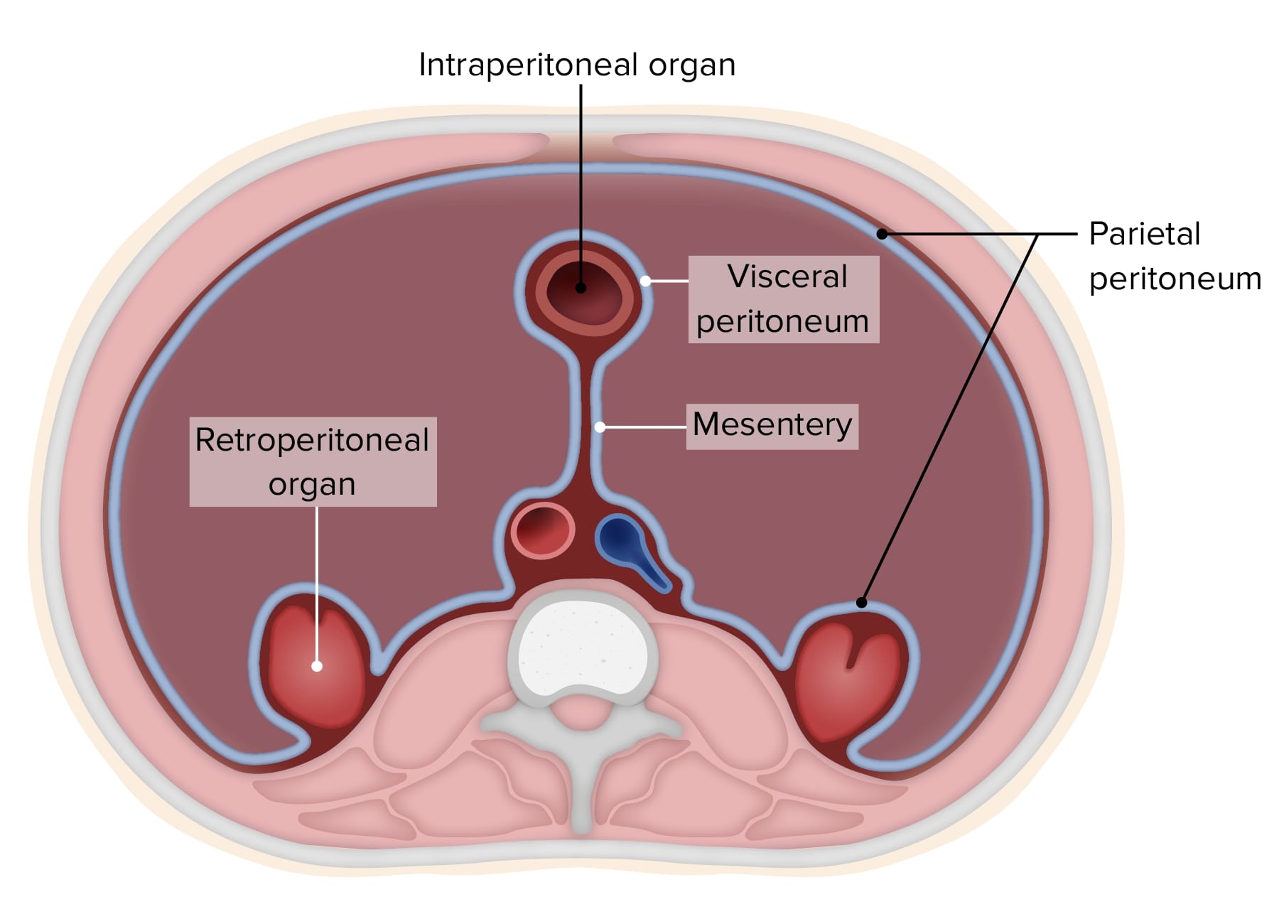
00:01 So now let's move on to another topic which we've spoken about quite a bit during the topics beforehand, but haven't really explained what it is. Let's talk about the peritoneum. So, the peritoneum is a membranous layer that really lines a lot of the abdomen. So let's have a look at the diagram to try and simplify, just understand the basic principles of what the peritoneum is. So let's just remind ourselves of this view is looking at a transverse section through the abdomen and at the bottom of the screen we've got the person's back. So it's if the person is lying on their bed and you're looking at that transverse dissection body through their feet. So here we can see the back is on the bottom of the screen and their anterior abdominal wall is pointing up towards the top of the screen. We can see that just in front of the spinal cord and the vertebral colon to the right hand side, we have our 2 kidneys and then essentially we have the aorta, the inferior vena cava. To the far right hand side, we have the ascending and descending colon and then we have the coils and coils of small intestine. 01:06 So that will help just to orientate ourselves because we'll look at this image a few times during the course of this video. So the peritoneum. The peritoneum is the innermost lining of the anterolateral abdominal wall. We've spoken about that a lot when we look to the anterolateral abdominal wall and hernias in the inguinal region It is a continuous layer though that extends over the posterior abdominal wall. So you can see it's lining all of the abdominal wall, but it also is invested around the individual organs. So it's covering a lot of the reservoir like the tubes of the gastrointestinal tract and also as we'll see to varying extents the accessory organs of digestion. The piece of peritoneum that is lining the body wall is known as parietal peritoneum. The continuous layer of peritoneum that's lining a piece of reservoir is known as visceral peritoneum, but these are the same sheath. 02:08 It is one continuous sheath of membrane which we call peritoneum. But when it's touching a piece of body wall, we call it parietal. And when it's touching a piece of reservoir, we call it visceral. So let's have a look at the peritoneal cavity. This is then the space that we find between the visceral and peritoneal and parietal layers of peritoneum. 02:33 So here we can see the peritoneal cavity, and it's filled with a very thin layer of fluid which helps to allow the various organs, the gastrointestinal tract, to move around the abdomen. So it has a very small layer of peritoneal fluid, and that helps to reduce friction. Here, we can see a piece of gastrointestinal tract. We can see that it's completely surrounded by a piece of peritoneum except for small beads that is called the mesentery. And that's the little connecting piece in yellow which we can see the peritoneum just being separated. Where we have this peritoneal layer completely covering the gastrointestinal tract. We call this an intraperitoneal organ. It's completely surrounded except for very small little root, and this is an intraperitoneal organ. If we indicate in here the kidney, what this is a retroperitoneal organ. The kidney lays on the posterior abdominal wall and essentially it just had a sheath of peritoneum layed over it. So it's retro, it's behind the peritoneum. So it's a retroperitoneal organ.
About the Lecture
The lecture Peritoneum: Overview by James Pickering, PhD is from the course Peritoneum and Peritoneal Cavity.
Included Quiz Questions
Which structure is posterior to the peritoneal cavity?
- Retroperitoneal cavity
- Greater sac
- Lesser sac
- Infracolic compartment
- Supracolic compartment
Customer reviews
5,0 of 5 stars
| 5 Stars |
|
5 |
| 4 Stars |
|
0 |
| 3 Stars |
|
0 |
| 2 Stars |
|
0 |
| 1 Star |
|
0 |


