Playlist
Show Playlist
Hide Playlist
Development of the Kidneys and the Adrenal Glands

-
Slides 08-48 Microscopic Development of the Kidney and Suprarenal Gland.pdf
-
Reference List Embryology.pdf
-
Download Lecture Overview
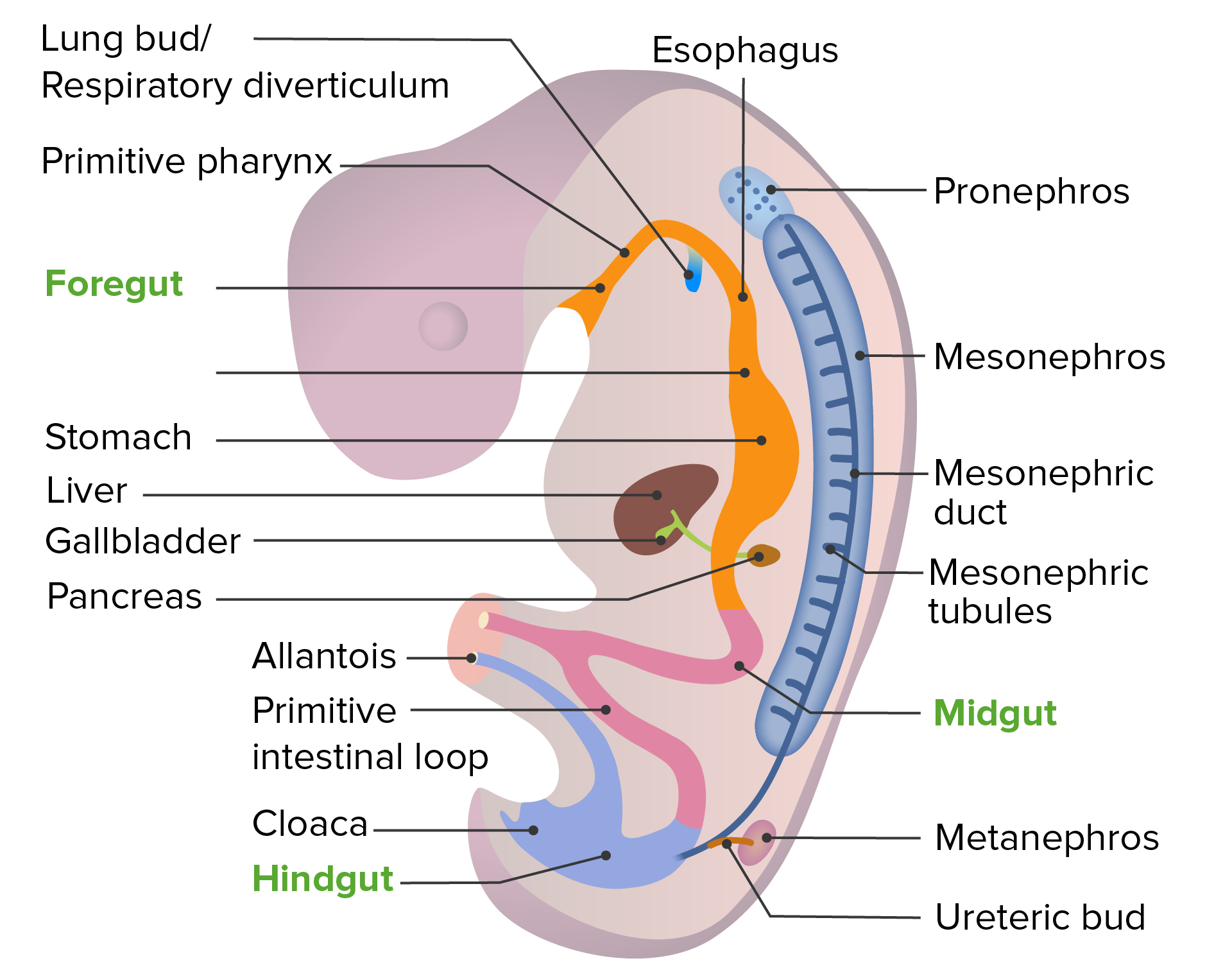
00:01 We will continue our discussion of renal development by looking at how the microscopic development of the metanephric kidney takes place which gives rise to the actual filtration system of the kidney. 00:12 We have two sets of kidneys formed during early life. 00:16 The mesonephros forms, develops a mesonephric duct that actually starts secreting urine into the urinary bladder. 00:24 So the tubules of the mesonephric duct associate with a glomerulus, filtration occurs into the renal or Bowman’s capsule, and urine is released to the bladder from the mesonephric kidney. 00:36 However, our body has decided that that wasn’t quite good enough and we have a ureteric bud come off of the mesonephric duct, induce formation of the final kidney or the metanephros which will then begin filtering urine and releasing it to the bladder. 00:53 The mesonephros dwindles away to pretty much nothing but its ductwork and its tubules stick around and associate with the nearby gonad. 01:02 Now, at this stage, the gonad is completely undifferentiated or indifferent. 01:06 It’s neither a testis nor an ovary yet but in the male, the mesonephric duct will stick around and form a continuous connection from the gonad, down into the urinary system. 01:18 We’ll follow that in an entirely separate talk but right now, we’re gonna instead move back to the ureteric bud and the metanephros and see how the filtration system of the kidney finally develops. 01:30 As the ureteric bud goes into the metanephros, it starts splitting in more and more finely woven little pathways that invade the metanephros. 01:42 In the process, it will create the renal pelvis, the major calyces, and even smaller level, the minor calyces, and an even smaller level, it will actually invade the substance of the metanephros and form the collecting ducts and the collecting tubule of the kidneys. 01:58 So here, we can see at the very, very fine magnification that collecting ducts have connecting tubules kind of budding off of them and associating with the tissue of the metanephros. 02:11 The metanephric vesicle is going to expand outward and develop a lumen, develop a little hollow space inside and at the same time, a hollow space or lumen is developing in the collecting ducts and collecting tubules. 02:25 Now, this vesicle of the metanephros that’s growing away from the ureteric buds collecting tubule is going to elongate and it’s gonna start snaking around and developing a loop. 02:37 So this S-shaped vesicle will eventually turn into the spiral and then, looped structure of the loop of Henle and the rest of the nephron. 02:49 So we have two embryonic origins for the urinary drainage apparatus. 02:54 The metanephros is giving rise to the nephron and then, the collecting system is coming from the ureteric bud. 03:01 But they form a continuous lumen that allows blood to be filtered at Bowman’s capsule and then, solutes, sugar, and other substances recovered from it before the urine is released into the collecting tubule, collecting duct, and then, into more and more large portions of the drainage apparatus. 03:22 That’s going to be the minor calyces, major calyces, renal pelvis, ureter, and finally, the urinary bladder. 03:30 Now, filtration of amniotic fluid actually does occur during life. 03:36 We breathe in and swallow amniotic fluid. 03:40 That fluid winds up in our tissues and our embryonic and fetal bodies filter that amniotic fluid and actually release urine back into the amniotic fluid. 03:52 Now, that sounds horrible. 03:53 The good news is the placenta does such a good job of filtering out all the waste products and other nasty things that are present in our body that the urine that’s excreted in the amniotic cavity is very, very non-threatening. 04:10 It doesn’t really do any damage. 04:11 It’s very much like amniotic fluid in, amniotic fluid out. 04:16 So we’re not polluting the amniotic sac much even though that process is occurring throughout fetal life. 04:22 Nearby, we have the suprarenal or adrenal glands forming and they come from two distinct sources. 04:30 Initially, we have the cortex migrate in from an area near the urogenital ridge and form a little C-shaped cup that associates with neural crest cells that are migrating in to form the adrenal medulla. 04:45 The portion that came from the cortex, pardon me, the portion that gives rise to the cortex is coming from near the urogenital ridge and it happens not just once, but twice. 04:55 There’s a fetal cortex that forms around the adrenal medulla and then, the adult cortex moves in and this finally creates the trilaminar appearance of the adrenal cortex and surrounding those neural crest cells, we’ll have the medulla. 05:12 So the medulla is derived from neural crest cells and they’re essentially going to release epinephrine and norepinephrine and they are essentially sympathetic nerve cells that instead of migrating into the gut to release norepinephrine and epinephrine at a synapse, are going to release it into our blood supply. 05:30 So just remember that the adrenal cortex is coming from an area near the urogenital ridge but the adrenal medulla is derived from neural crest cells. 05:42 Now, things that can go wrong with kidney development include oligohydramnios. 05:48 Now, oligohydramnios essentially means too little amniotic fluid and problems with the kidneys are only one cause of oligohydramnios. 05:57 Leakage of amniotic fluid can also cause the same problem but renal agenesis or blockage of the urinary drainage system is a fairly common cause of it. 06:09 And if you look at the picture here, you can note that these kidneys don’t exactly look healthy but in particular, you can see that the ureter on the one on the left side of the image is pinched. 06:18 Failure of urine to be filtered because there’s no kidney or failure of urine to exit the kidney can cause Potter sequence. 06:27 Potter sequence is a problem that develops from oligohydramnios, too little amniotic fluid. 06:33 The amniotic fluid acts like a little buoyant kind of support for the developing body. 06:39 We’re floating around in the amniotic fluid and it keeps us relatively cushioned. 06:43 If there’s too little amniotic fluid, then the fetus is being kind of pushed down by gravity. 06:50 This can cause limb defects, hip dislocation, and also, facial defects because the face is pushed up against the wall of the uterus instead of being able to kind of float relatively free. 07:01 So Potter sequence is a sequence of events that follow from lack of amniotic fluid. 07:07 Thank you very much for your attention and I’ll see you in our next talk.
About the Lecture
The lecture Development of the Kidneys and the Adrenal Glands by Peter Ward, PhD is from the course Development of the Abdominopelvic Region.
Included Quiz Questions
The metanephric S-shaped vesicle will eventually become which of the following?
- Nephron
- Metanephric cap
- Collecting tubule
- Metanephric vesicle
- Pear-shaped vesicle
Which of the following structure is the precursor to the collecting tubules?
- Ureteric bud
- Metanephric cap
- Metanephric S-shaped vesicle
- Pronephros
- Metanephric pear-shaped vesicle
Which of the following best describes the development of the suprarenal gland?
- Two waves of cortex come in from around the urogenital ridge and migrating cells from the neural crest invade this developing cortex to form the medulla.
- One wave of cortex comes in from the urogenital ridge and surrounds the neural crest cells, which ultimately form the medulla.
- Two waves of neural crest cells come in to become the cortex and surround the migrating urogenital ridge cells that will ultimately become the medulla.
- One wave of neural crest cells come in from the urogenital ridge to become the medulla and subsequently, cortical migration occurs from cells in the urogenital ridge to become the cortex.
- Three waves of cells migrate from the urogenital ridge to form the trilaminar suprarenal gland, including two layers of cortex and one layer of the medulla.
Which of the following is not associated with Potter sequence?
- Failure of the neural crest cells to migrate into and become the medulla of the suprarenal gland
- Oligohydramnios
- Facial flattening
- Limb malformations due to long-term lack of buoyancy for the developing fetus
- Failure of lung development
Author of lecture Development of the Kidneys and the Adrenal Glands

Peter Ward, PhD
Customer reviews
5,0 of 5 stars
| 5 Stars |
|
5 |
| 4 Stars |
|
0 |
| 3 Stars |
|
0 |
| 2 Stars |
|
0 |
| 1 Star |
|
0 |
