Playlist
Show Playlist
Hide Playlist
Ovaries and Paramesonephric Ducts

-
Slides 08-51 Ovaries and Paramesonephric Ducts.pdf
-
Reference List Embryology.pdf
-
Download Lecture Overview
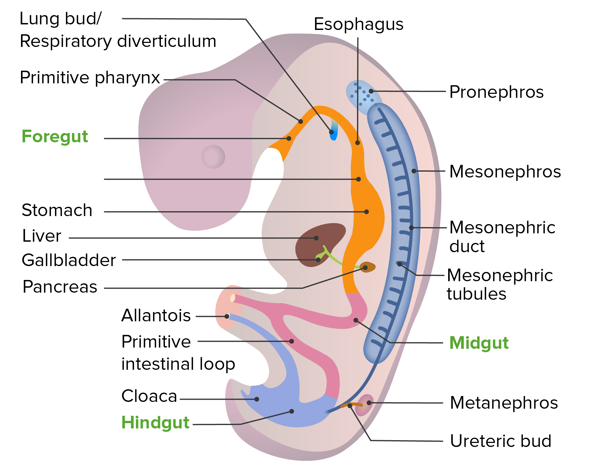
00:01 In this talk on reproductive development, we will follow the structures that form specific female internal genitalia and organs. 00:09 Typically, the chromosomes that are present in a person’s body will kick start either male development if there are XY chromosomes present or female development if XX chromosomes are present or no specific male signals are released by the body. 00:27 So female development will happen in the absence of a Y chromosome even if there are some other chromosomal abnormalities that we’ll discuss later. 00:35 In an XX individual, the primitive sex cords that have grown in from the genital ridge will regress and there will instead be cortical cords that grow in from the epithelium on the outside of the intermediate mesoderm. 00:50 These will associate with the primordial germ cells that have migrated into the area from the yolk sac a little bit earlier. 00:57 These will then break lose and the cortical cord cells will associate with the primordial germ cells forming primordial follicles. 01:08 High levels of estrogen not only allow this set of circumstances to develop but also, cause the Wolffian or mesonephric ducts to regress but maintain the structures that are nearby, the paramesonephric ducts. 01:23 So near the ovary, the paramesonephric ducts open and will be staying in close association with the ovary even though they never actually invade the ovary. 01:34 They just associate with it nearby. 01:36 The paramesonephric ducts on the left and right will come together to form the uterine body, cervix, and the superior most portion of the vagina. 01:45 Small remnants of the disintegrating mesonephric duct can sometimes be found nearby in the mature ovary and these go by the excellent names of epoophoron and paraoophoron and occasionally, can be seen flanking the uterine body and are thereafter known as Gartner’s duct cysts or just Gartner’s cysts. 02:08 These are small remnants of the mesonephric duct and are typically clinically unimportant unless they develop problems that lead to cancer of those specific cells. 02:18 Now, we’ve mentioned that the paramesonephric ducts have come together to form a common chamber that will be the body of the uterus. 02:27 These associate with the urogenital sinus but instead of simply opening into the urogenital sinus, something a little more complex happens. 02:36 Growths off of the urogenital sinus called sinovaginal bulbs grow upward to meet the paramesonephric ducts, thicken and actually form a solid plate of cells called the vaginal plate. 02:50 Now, overtime, the vaginal plate will develop a lumen and connect the uterus, cervix, and vagina to the opening of the vestibule. 03:01 There will be a thin membrane there marking the location of where the vaginal plate met the urogenital sinus and that is going to be the hymen. 03:08 We can here see a sagittal view of the same process. 03:12 The paramesonephric ducts have fused to create the uterine body which is associated with the urogenital sinus, the sinovaginal bulbs and then, the vaginal plate enlarge connecting the paramesonephric ducts to the vestibule. 03:27 As a lumen develops in the vaginal plate, we’ll have a continuous chamber going from the vestibule, into the vagina, to the uterus, and thereafter, the oviducts. 03:38 However, the place where the vestibule and sinovaginal bulbs meet is marked by the hymen. 03:44 Things that can go wrong in the process involve Turner syndrome which is when only a single X chromosome is present or if you have significant deletions or damage to an X chromosome, Turner syndrome can still develop. 03:59 People with Turner syndrome typically are of short stature and have a webbed neck, sometimes extending from the base of the ears, all the way out to the tips of the shoulders, lymphedema, or swelling in the limbs, a wide chest with widely spaced nipples, and amenorrhea, failure to generate regular menstrual periods. 04:17 They typically have no intellectual disabilities and the ovaries are often absent or sometimes, regressed into a little fibrous cord sometimes called streak ovaries. 04:28 Now, as the paramesonephric ducts fuse to create the uterine body and then cervix, and proximal portion of the vagina, a few problems can develop if they don’t fuse or don’t form properly. 04:40 If they stay separate, you can actually have a double uterus with a septum running down the cervix and vagina. 04:49 So functionally, two separate openings of the vagina into the vestibule, leading to two separate uterine cavities and two separate oviducts. 04:58 Less severe would be of no septum present in the vagina but a clear separation of the uterus. 05:06 This can occur with completely separate uterine bodies or a small common chamber with two uterine horns extending from it. 05:16 So here, and here, we have an example of a bicornate uterus. 05:21 One with a common chamber, one without. 05:24 Less severe is going to be the presence of a membranous septum marking the place where the two mesonephric ducts came together and instead of rupturing to create a single uterine cavity. 05:35 There’s only a small membrane separating the two halves of it. 05:39 These conditions may be completely unknown to the affected woman until attempting to get pregnant. 05:45 At which point, there may be difficulties with implantation and growth of the fetus in the womb. 05:51 If the paramesonephric ducts fail to form or form in a slightly atrophic way, you can have a rudimentary horn of the paramesonephric duct leading to a small and slightly kind of dwindled version of the uterus on that side but the ovary will likely be present. 06:10 It’s simply not able to shed eggs into the oviduct nearby. 06:15 Failure of a paramesonephric duct to form altogether on one side results in uterine atresia. 06:22 Now, this can happen unilaterally or bilaterally. 06:26 In which case, the vagina will form from the sinovaginal bulbs but it will not connect to a uterus. 06:32 Vaginal atresia itself can happen if even those sinovaginal bulbs fail to form or the vaginal plate does not open up and create a lumen. 06:43 And occasionally, you can have vaginal atresia with a completely normal uterus on the other side, just no canal in between. 06:51 Now, the place where the sinovaginal bulbs and vaginal plate meet the urogenital sinus, we have formation of the hymen. 07:00 Normally, the hymen will rupture over time, typically, around puberty. 07:04 But if the hymen either does not rupture or has a very small opening, we can have incomplete perforation of the hymen, this can be medium or very small. 07:15 We can have a septation of the hymen where strips of tissue are covering the opening of the vagina. 07:22 There can be cribriform hymen where there are multiple small openings. 07:27 And there can be completely imperforate hymen in which case, none of the secretions of the vagina, cervix, and uterus are able to be released, and this may need to be fixed surgically. 07:39 Okay, thank you for your attention and I’ll see you in our next talk.
About the Lecture
The lecture Ovaries and Paramesonephric Ducts by Peter Ward, PhD is from the course Development of the Abdominopelvic Region. It contains the following chapters:
- Ovaries and Paramesonephric Ducts
- Uterine Defects
Included Quiz Questions
High levels of estrogen lead to which of the following developmental event?
- The regression of mesonephric ducts
- The regression of paramesonephric ducts
- The development of the mesonephric ducts
- The activation of the testis-determining factor on the Y chromosome
- The development of a bicornate uterus
Which of the following is false of the vaginal plate?
- It develops into the uterus.
- It develops into the vagina.
- It creates vaginal hymen.
- It connects the uterus to the vestibule.
Which of the following statements about Turner syndrome is false?
- It typically results in intellectual disability.
- It occurs when a female has only a single X chromosome or significant deletions to one X chromosome.
- Ovaries in a Turner syndrome patient are usually absent or fibrous, called 'streak' ovaries.
- Individuals with Turner syndrome typically are born with a 'webbed' neck.
- It occurs in X monosomic mosaic individuals.
Failure of the left and right paramesonephric ducts to fuse can result in all but which of the following?
- Rudimentary uterine horn
- Double uterus with vaginal septum
- Double uterus without vaginal septum
- Bicornate uterus
- Uterine septum
Author of lecture Ovaries and Paramesonephric Ducts

Peter Ward, PhD
Customer reviews
4,5 of 5 stars
| 5 Stars |
|
1 |
| 4 Stars |
|
1 |
| 3 Stars |
|
0 |
| 2 Stars |
|
0 |
| 1 Star |
|
0 |
he is the only lecturer who helped me to understand the embryology!!! he is gold!!!
As you mention words/features, would be nice if the words on the screen were highlighted on the images. Hard to find them in the midst of listening.
