Playlist
Show Playlist
Hide Playlist
Osteopathic Medicine Approach: Foot and Ankle

-
Slides Osteopathic Diagnosis of the Ankle and Foot Region.pdf
-
Reference List Osteopathic Manipulative Medicine.pdf
-
Download Lecture Overview
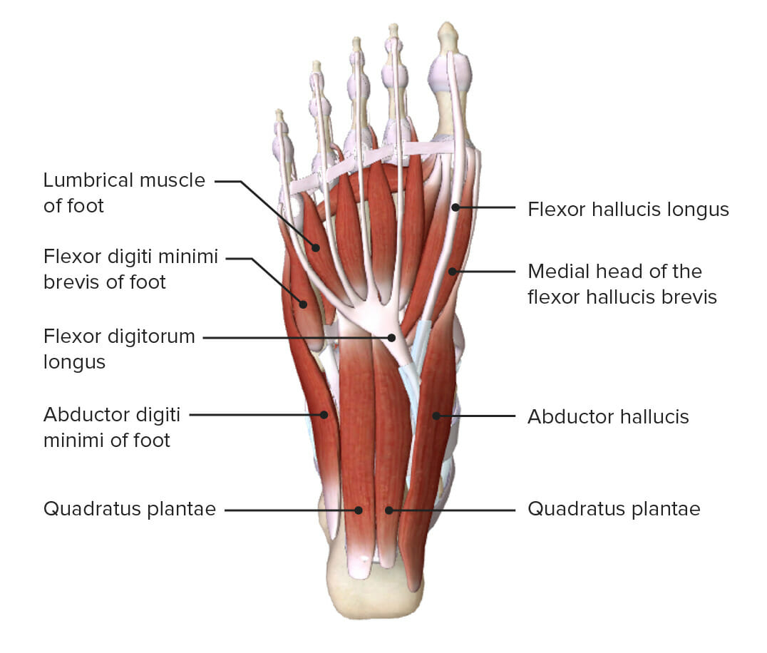
00:01 We're gonna talk about how do you approach a problem in the foot and ankle from an osteopathic medicine perspective. 00:06 This is gonna be heavy on anatomy, heavy on learning the spaces, the attachments and the muscle functions of the foot as well as the joint motions. 00:16 We'll talk about different tests you can do and also have separate videos for those tests. 00:22 And we'll talk a little bit about ankle sprains as the most common issue noted in the ankle. 00:27 So when you want to examine the ankle, you want to inspect one joint above, one joint below. 00:33 You want to make sure the motion of the ankle, the function of the ankle is smooth and fluid. 00:38 You want to make sure that the foot and the ankle during the weight bearing phase of walking, are fluid as well because that's where an injury may occur, a fall may occur or other issues. 00:50 You also want to look for skin changes, changes in color and changes that may signal other issues. 00:59 And you want to look for any swelling or signs of trauma. 01:02 So let's start with the anatomy of the foot. 01:04 When you look at the foot and you look at the medial aspect, one of the bones that we're gonna be very focused on is the navicular bone. 01:13 We don't want to miss a fracture and it often feels like an ankle sprain but the navicular bone can be broken The navicular bone articulates with 5 other bones. 01:24 It has three cuneiforms and a cuboid bone all attaching it. 01:29 So, we are gonna touch it, make sure we feel it and understand the motion that you can induce. 01:35 We also worry about a septic necrosis of the navicular bone occuring, especially in children. 01:43 So the navicular and the head of the talus will touch. 01:47 The talar head is immediately proximal to the navicular bone, closer to the body And these are the bones that are gonna cause inversion and eversion of the forefoot. 01:59 So it's gonna create motion in the foot that's gonna happen only in that bone. 02:04 It's not gonna be a great motion but it is very important motion. 02:09 We're gonna talk a little about the medial malleolus, the inside of the ankle that sticks out. 02:14 It's part of the talus connection, it creates the ankle mortise and it comes from the tibia. 02:22 So it's an important bone. 02:25 Under that bone you're gonna have the sustentaculum tali,which is a space. 02:31 It is difficult to palpate, it's there, It will support the talus and serve as an attachment for the spring ligament. 02:40 So these are important areas to be aware of because there is motion that's gonna be occuring in this area as well, and you may have some swelling and tenderness. 02:49 The 5th metatarsal bone and the 5th metatarsophalangeal joint are important areas to explore, to examine, to touch, to make sure enough there isn't any tenderness It's also a frequently broken area. 03:07 It's the lateral side of the ball of the foot towards the back of it and it's along the lateral side of the fifth metatarsal and the styloid process there can often be palpated And if you palpate it too much, it is somewhat tender. 03:21 When you look at the lateral aspect of the foot, there are muscles there that are gonna cause the eversion as well with the peroneous brevis, one that you can feel and is important to look out when you're doing the exam. 03:34 There's a groove located just behind the cuboid bone that's created by the peroneus longus and peroneus brevis that you want to feel and examine because the motion is going to be causing in the foot and allowing the foot to plantarflex, to push down into the ground. 03:54 The sinus tarsi area is just anterior to the lateral malleolus. 03:59 So if you go to the lateral malleolus, the little bone out cropping on the outside and anteriorly, you'll see the sinus tarsi region, This is a soft tissue depression between the bones and is also where the extensor digitorum muscle will go and there's an overlying fat pad that can be touched as well. 04:23 The dome of the talus is also an important bone. 04:26 It can be felt in inverting and plantarflexing the foot, and helps with motion, will be very tender at times if the motion is abnormal and it could be palpated more laterally than medially. 04:41 So the dome of the talus is an important area to be aware of. 04:45 The medial tubercle of the calcaneus is something you hold on to a lot when you're doing manipulation, you use it to stabilize the foot. 04:52 It's on the medial plantar surface of the calcaneus and it gives you a good grip in stabilizing the foot. 04:58 There are a lot of muscle attachment here particularly at ABductors. 05:03 The abductor hallucis muscle comes medially and the flexor digitorium brevis muscle will also attach and you'll have the plantar aponeurosis that may cause plantar fasciatis may pull the arch and it's something we'll talk about in just a few minutes when we talk about the arches. 05:19 And this is also important because when you walk, this is where heel strke occurs and the first touch of the ground is, and you may notice tenderness or early contraction of the toes. 05:33 So these are just things that all come together you should be aware of. 05:37 When you look at the plantar surface, you'll also notice there are some sesamoid bones Sesamoid bones are small round bones that are embedded in a ligament or in a fascia. 05:49 You'll find sesamoid bones typically by the first metatarsal, the base on the first toe just under the big toe. 05:58 You'll have at least two sesamoid bones within the flexor hallucis brevis tendon which help with motion and also allows for some distribution of the body weight when you're walking by displacing the force amongst more than just that one area so you don't develop more osteoarthritis or risk breaking something or having overuse injuries. 06:21 We talk about arches, we talk about the medial and lateral longitudinal arches and the the anterior metatarsal arch creating the biggest portion of the foot arch that allows you to walk comfortably gives you a sponginess to your walk and gives you comfort You can also think of the posterior metatarsal arch which is smaller and the tarsal arch which also is in existence. 06:43 When we look at soft tissue landmarks, the navicular tubercle and the talar head are important because while they lack boney support, they do support some of the muscles by attaching, by being a tendon attachment and giving you the spring ligament and the sustentaculum tali area So it is just an important part to notice. 07:06 When people have flat feet, the navicular tubercle will displace medially and plantarward. 07:15 So you're gonna have the foot bent upward toward the body. 07:19 The deltoid ligament is a medial ligament, It is a very strong ligament made up of multiple pieces and this is one that can be torn during an ankle sprain It has the tibialis posterior tendon running through it and the flexor digitorum tendon running just behind it as well. 07:40 It's important to be aware of the deltoid ligament because you're gonna palpate a lot of these You're gonna learn what the normal size is, what the normal distribution is and when you feel a piece of it gone, or a piece of it pulled off, you know there's gonna be an ankle sprain. 07:54 The flexor hallucis longest tendon is also another one. 07:58 It's right behind the ankle joint. 08:00 The ankle mortise is the tibia, the fibula coming together form a very strong hinge joint of the foot and the flexus hallucis longus which moves the toe is going to be behind it and you can't palpate it because it's deep to the other muscles. 08:18 The posterior tibial muscle and the tibial nerve are also in the back portion of the foot between the tendons and the flexor digitorum longus and flexor hallucis longus muscle. 08:31 And these are the ones inducing movement to the foot With the neuromuscular bundle bound by the tarsal tunnel. 08:38 So it's gonna be deeper and somewhat protected. 08:42 The anterior talofibular ligament is the lateral ligament, it is frequently sprained or strained. 08:51 It's part of 3 ligament that are that attaching the tibia to the foot and calcaneofibular and the posterior talofibular ligament are the other two. 09:02 The posterior talofibular ligament is a pretty strong one and usually not torn. 09:06 It's usually anterior talofib and pieces of the calcaneo fibular ligaments Other soft tissue landmarks to be aware of are the proneus longus and brevis tendons and these are gonna be seen on the dorsal aspect of the foot. 09:23 They go just behind the lateral malleolus and they're primarily foot everters and they sometimes assist in plantarflexion as well, pushing the foot down. 09:36 This is something you can also snap on occasion and you're gonna hear people and what sometimes sounds like a click or a pop but it's just a snapping tendon when it goes over a tuberousity, a little outcropping of bone that the tendon can snap over. 09:53 The sinus tarsi is another soft tissue landmark on the top of the tarsal bone. 10:01 It's affected by ankle sprains in that some of the swelling goes there, some of the pain and the difficulty in walking arises from there. 10:09 There's a deep tenderness when you palpate it and they can sometimes confuse people into thinking that maybe a break because if you look at the Ottawa ankle rules, if you have tenderness within 6 inches up of the malleolus, you need to get an x-ray This may confuse you of the swelling that extends down and into this area. 10:28 The calcaneus again, the large bone in the back with achilles tendon inserts and the gastrocnemius will assist with motion, very strong tendon, can pop or snap with severe pressure or can be cut but it is easily reattached surgically When that does happen, you'll notice a positive Thompson's test which is when you squeeze the gastrocnemius and the foot doesn't move, will tell you there's been an interruption in the gastrocnemius tendon. 11:00 I just want to mention the retrocalcaneal bursa because bursas are important at easing motion. 11:06 This is the area between the anterior surface of the achilles tendon and the superior angle of the calcaneous. 11:12 and then there's the calcaneal bursa, another bursa which follows the insertion of the achilles tendon over that region.
About the Lecture
The lecture Osteopathic Medicine Approach: Foot and Ankle by Tyler Cymet, DO, FACOFP is from the course Osteopathic Diagnosis of the Ankle and Foot Region. It contains the following chapters:
- Osteopathic Medicine Approach – Foot and Ankle
- Arches of the Foot
- Soft-tissue Landmarks
Included Quiz Questions
Which of the following is the anatomical location of the sinus tarsi?
- Anterior to lateral malleolus, between the talus and calcaneus
- Posterior to the lateral malleolus
- Anterior to the medial malleolus
- Anterior between the talus and navicular
What group of ligaments make up the lateral ligament of the ankle?
- Anterior talofibular ligament, calcaneofibular ligament, posterior talofibular ligament
- Anterior talofibular ligament, posterior talofibular ligament and deltoid ligament
- Anterior tibiotalar ligament, tibiocalcaneal ligament, posterior talofibular ligament and tibionavicular ligament
- Anterior talofibular ligament, tibiocalcaneal ligament, posterior tibiotalar ligament and tibionavicular ligament
What anatomical structure is the distal insertion of the Achilles tendon?
- Calcaneus bone
- Navicular bone
- Talus bone
- Cuboid bones
Author of lecture Osteopathic Medicine Approach: Foot and Ankle

Tyler Cymet, DO, FACOFP
Customer reviews
5,0 of 5 stars
| 5 Stars |
|
5 |
| 4 Stars |
|
0 |
| 3 Stars |
|
0 |
| 2 Stars |
|
0 |
| 1 Star |
|
0 |
