Playlist
Show Playlist
Hide Playlist
Neurocysticercosis: Life Cycle, Presentation, and Disease Stages
-
Slides Other CNS Infections.pdf
-
Download Lecture Overview
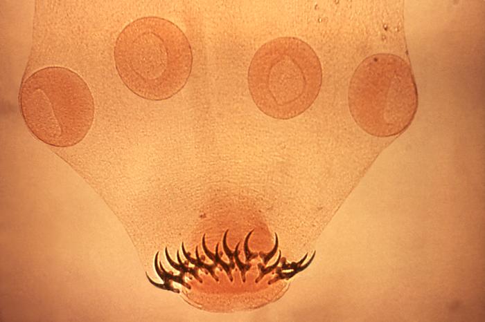
00:00 What's the life cycle of this organism? How does it work, how does it live, and how does it infect humans? Well, typically we think of the life cycle starting in pigs. Pigs ingest the eggs and those eggs develop into a cysticerci, which lodges or encyst in the muscle of pork and humans eat that meat. When humans eat the meat, the cysticerci arrive into the stomach and can grow and develop into the adult. The adult is called taenia solium and this arrives in the stomach of humans and the eggs are released in the feces and that cycle continues and that's the normal life cycle for this organism. In certain situations, we see an aberrant life cycle particularly if there is a problem with handling. There can be fecal oral transmission of the eggs to humans and this can occur with improper handling. Humans ingest the eggs, those eggs then develop into the cysticerci which encyst within the brain and the muscle and this leads to a symptomatic disease in humans. This is an important cause of new onset epilepsy and I'd like you to remember this graph not for all the specific details, but how common new onset epilepsy is in patients with neurocysticercosis. Over 60% of patients who have this infection will present with epilepsy. We see other things, intracranial hemorrhage, motor dysfunction, meningismus, dementia or cognitive dysfunction, psychotic syndrome, strokes and other things but the vast majority of patients present with new onset epilepsy. So what I want you to take away from this is a patient who is from or has travelled to an area endemic to neurocysticercosis and presents with new onset epilepsy should undergo imaging evaluation and consideration of this infection. Just as neurocysticercosis or the cysticerci have a typical life cycle, we also see a life cycle, a natural history of its development within the brain and that follows 4 stages and I'd like you to know of not all the details about but know of these 4 stages. There is the vesicular stage, the colloidal stage, the granular stage, and the calcified stage. And what's happening is the organism is growing, is becoming inflamed and recognized by the immune system, and then dies over time. The vesicular stage is the asymptomatic stage where this cyst lies dormant and is not seen or reacted to the immune system. In the colloidal stage, we see a robust and vigorous inflammatory response as the immune system attacks this organism and the organism degenerates. The granular stage is the physical degeneration of this parasite which then dies and it reaches the calcified stage and is no longer at risk for active infection within the brain and will calcify. So let's look at each of those in greater detail and focus on what's going on, what symptoms we may see in patients, and what the imaging looks like because imaging is the critical way we evaluate neurocysticercosis. So the vesicular stage, this is where the cysticercus enters the brain, there is a clear vesicular fluid and normal invaginated scolex, that's the mouth of this parasite. Typically, patients are asymptomatic, this is lying dormant in the brain not causing problems. There is scarce perilesional inflammation, this looks like a hole with a dot. So on a CT scan, you see a hole or a cyst. There is no enhancement around it, there is no edema around it, and this may lie dormant for years and not cause any problems. Over time, the immune system may start to recognize this parasite for reasons that are not clear and the timing really varies by patient and situation. 03:52 The parasite will enter the colloidal stage, the fluid will become turbid, the scolex undergoes hyaline degeneration, and at this point there is a vigorous and robust immune response against this parasite. In this stage, we see prominent symptoms. There is an intense mononuclear inflammation around this lesion, there is a capsule that becomes inflammatory and inflamed and enhances with contrast and patients will develop focal deficits at the area of this lesion during the colloidal stage. On imaging, contrast is critical and here we see an axial T1 weighted post contrast imaging showing surrounding enhancement gadolinium enhancement with some edema, some dark edema surrounding this lesion as it's undergoing active degeneration and immune attack. After the colloidal stage, the cyst degenerates and we enter the granular stage. The scolex transforms into granules. The cysticercus is no longer viable, it will not continue to grow or change over time, it's degenerating. And we see that, we see astrocytic gliosis, scarring of the brain, nodular contrast enhancement though there is still some inflammatory gliotic change occurring but no longer edema on the brain and typically minimal symptoms. And you can see that here by this enhancing nodular lesion on this axial image. Ultimately, the parasite dies and enters the calcified stage and during that stage there is intense gliosis. This is a scar on the brain that will never continue to infect the brain or develop an inflammatory response, what's done is done and what we're left with on imaging is a small calcified area.
About the Lecture
The lecture Neurocysticercosis: Life Cycle, Presentation, and Disease Stages by Roy Strowd, MD is from the course CNS Infections.
Included Quiz Questions
What is the most common clinical manifestation of neurocysticercosis?
- Seizure
- Meningismus
- Fever
- Change in behavior
- Motor signs
Which disease stage is characterized by a robust inflammation against the causative parasite?
- Colloidal stage
- Vesicular stage
- Viable stage
- Granular stage
- Calcified stage
What imaging findings would be expected during the granular stage of neurocysticercosis?
- Nodular contrast enhancement
- Hole with a dot
- Perilesional edema
- Ring-enhancing capsule
- Multiple intracranial calcification
Customer reviews
5,0 of 5 stars
| 5 Stars |
|
1 |
| 4 Stars |
|
0 |
| 3 Stars |
|
0 |
| 2 Stars |
|
0 |
| 1 Star |
|
0 |
Me gustó mucho, mañana tengo una prueba sobre el tema y me ayudó a entenderlo


