Playlist
Show Playlist
Hide Playlist
Neurocysticercosis: Introduction

-
Slides Other CNS Infections.pdf
-
Download Lecture Overview
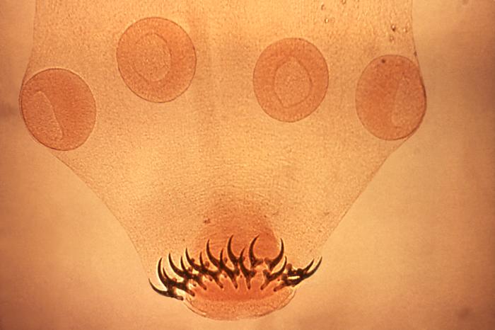
00:00 Now let's move to our 3rd type of opportunistic or exposure-related infections and talk about neurocysticercosis. This is an infection that's not seen typically in opportunistic host, patients who may be immunocompromised, but is seen in certain exposures, travel or occupational risk. 00:20 This is the most common parasitic infection in the brain. This organism is endemic to certain areas of the world and we think of Mexico, Central America, South America, Asia, and certain areas of Africa. Patients who are from or have travelled to those areas and develop a presentation concerning for CNS infection should be evaluated for this process. Imaging is important and we're going to walk through some of the imaging findings in these patients. 00:46 But you can see here multiple cysts it's called neurocysticercosis, multiple cysts throughout the bilateral hemispheres in this patient who has a neurocysticercosis infection. The typical clinical manifestation, patients present with seizures. So this is a little bit different in some of the other CNS infections we have talked about. We talked about headache pointing us to the brain, fever pointing us to infection, and then we think of meningismus for meningitis, altered mental status for encephalitis, and focal neurologic deficit for cerebritis, these patients often present without any of those findings and with first time seizure with or without a focal neurologic deficit. Headache can be seen, confusion can occur particularly in patients who have CSF dissemination of this organism. Ataxia can occur if the cerebellum is involved and we can see meningismus particularly if there is a secondary inflammatory meningitis from CSF spill over infection. Hydrocephalus or increased intracranial pressure can be seen in 25% of patients, but is not particularly common. And we diagnose this in patients who have at-risk travel, present with a chronic course or new onset seizure with imaging and typically we think of either CT or MRI.
About the Lecture
The lecture Neurocysticercosis: Introduction by Roy Strowd, MD is from the course CNS Infections.
Included Quiz Questions
Which of the following countries has a high incidence of neurocysticercosis?
- Mexico
- Australia
- New Zealand
- Canada
- Germany
Author of lecture Neurocysticercosis: Introduction

Roy Strowd, MD
Customer reviews
5,0 of 5 stars
| 5 Stars |
|
5 |
| 4 Stars |
|
0 |
| 3 Stars |
|
0 |
| 2 Stars |
|
0 |
| 1 Star |
|
0 |
