Playlist
Show Playlist
Hide Playlist
Nasal Cavity and Paranasal Sinuses

-
Slides Anatomy Nasal Cavity Paranasal Sinuses.pdf
-
Reference List Anatomy.pdf
-
Download Lecture Overview
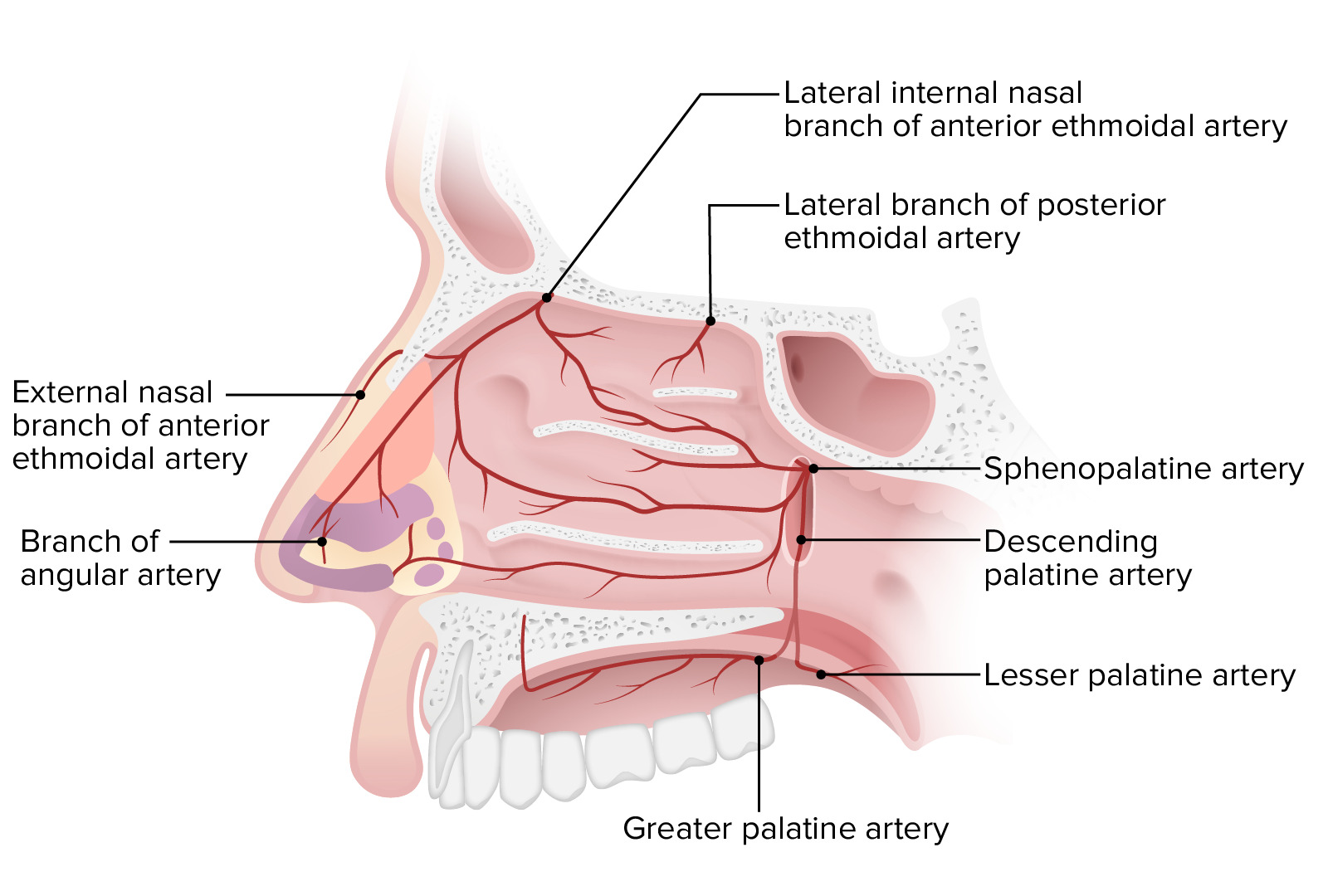
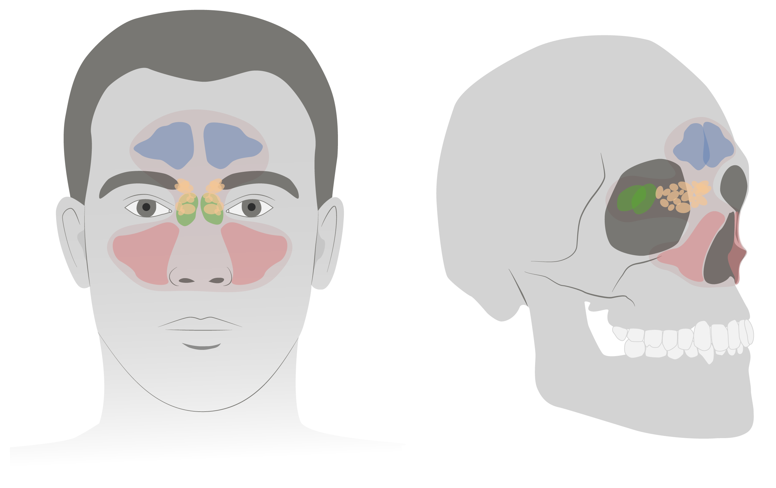
00:01 The next area we're going to look at is the nasal cavity and its associated paranasal sinuses. 00:09 We'll start externally by looking at the external nose. 00:14 Here we see the root, bridge, dorsum of the nose. 00:20 And then the anterior most portion is called the apex. 00:25 Laterally we have the ala or ala nasi and the opening is called the nostril. 00:34 We take off the skin, we can see the nasal bone which attaches to the nasal cartilages most anteriorly which gives the external nose its flexibility. 00:46 Let's look at the cavities of the nose or the nasal cavities. 00:52 In the midline, we have the medial wall and this is the nasal septum that separates the cavity into a left and right nasal cavity. 01:01 Here we have the lateral wall, the roof and the floor of the nasal cavity. 01:08 The posterior border of the nasal cavity something called the choana. 01:12 Beyond that we're going to have the nasal pharynx. 01:16 So let's look at this lateral wall of the nasal cavity. 01:20 We have the maxilla or the medial surface of the maxilla. 01:24 We have a little bit of the lacrimal bone. 01:29 We also have the ethmoid bone because the ethmoid bone is where we have the superior and middle concha as well as the ethmoid labyrinth. 01:39 The inferior nasal concha on the other hand is not part of the ethmoid. 01:44 It's actually its own bone. 01:48 Just posterior to that, we have the perpendicular plate of the palatine bone. 01:53 And beyond that, we have the medial plate of the pterygoid process of the sphenoid bone. 02:01 In terms of the roof of the nasal cavity, we have a little bit of the nasal bone anteriorly, the nasal spine of the frontal bone and then the cribriform plate of the ethmoid bone which is going to be very important for the passageway of olfactory nerves. 02:19 Posterior Lee we have the anterior surface of the sphenoid bone or we also have the openings to the sphenoidal sinus. 02:27 The floor of the nasal cavity is actually the palate. 02:31 So we see the palatine process of the maxilla, anteriorly. 02:35 And the horizontal plate of the palatine bone, posteriorly. 02:41 The medial wall that separates the nasal cavity and the left and right is the nasal septum. 02:47 And here we have the perpendicular plate of the ethmoid bone as well as the vomer and then the septal cartilage more anteriorly. 02:59 If we swing around to a posterior point of view, we see the posterior borders which are the nasal choanae. 03:08 And inferiorly, we again have the horizontal plane of the palatine bone. 03:13 Mediately, the posterior bodies of the vomer which make up part of the nasal septum. 03:20 Laterally, the medial plates of the pterygoid process and the roof again parts of the vomer in sphenoid bone. 03:30 If we were to take a cross section view, through the nasal cavities, we would see that there's some other spaces that are actually associated with the nasal cavity. 03:39 We call these the paranasal sinuses. 03:43 So here we see the septum in the midline and then the nasal concha on the lateral wall of the nasal cavity. 03:52 Here we have the orbit and then we start to see the paranasal sinuses. 03:58 We have some in the frontal bone called the frontal sinus, some of the ethmoid bone called the ethmoid sinus sometimes called air cells. 04:08 And then we have maxillary sinuses and the maxillary bone. 04:15 These parent nasal sinuses, we don't really know what they're doing there. 04:20 They might be making the skull lighter, they might be adding resonance to the voice. 04:25 But either way, the important thing is that they all communicate with the nasal cavity and as such, infections can communicate as well and you can get sinusitis. 04:37 So, these sinuses, all have some sort of connection. 04:42 For example, in the sagittal view, we see the sphenoid sinus entering the nasal cavity through the sphenoethmoidal recess. 04:52 We also have the posterior ethmoid sinus training through the superior meatus which is an opening just below the superior nasal concha. 05:03 The middle meatus, which is the opening just below the middle concha is where we have drainage of the frontal sinus, as well as the anterior and middle ethmoidal sinuses. 05:14 It's also where we have the maxillary sinus draining. 05:18 So we have multiple sinuses draining in this middle meatus. 05:22 In the inferior meatus or the opening below the inferior concha, this is where we have drainage of the nasolacrimal duct. 05:30 So this is essentially where tears end up draining. 05:34 And that's why if you've ever cried for a long period of time, it seems like your nose might be getting runny. 05:39 Well that's because of this drainage into the nasal cavity via the nasolacrimal duct.
About the Lecture
The lecture Nasal Cavity and Paranasal Sinuses by Darren Salmi, MD, MS is from the course Upper Aerodigestive Tract.
Included Quiz Questions
What is the most anterior part of the nose?
- Apex
- Root
- Bridge
- Dorsum
- Septum
What is the posterior border of the nasal cavity?
- Choana
- Maxilla
- Lacrimal bone
- Ethmoid bone
- Septum
What are the components of the roof of the nasal cavity? Select all that apply.
- Nasal bone
- Nasal spine of the frontal bone
- Cribriform plate of the ethmoid
- Lacrimal bone
- Maxilla
What sinus communicates with the nasal cavity via the sphenoethmoidal recess?
- Sphenoid
- Frontal
- Maxillary
- Ethmoidal
- Nasolacrimal
Author of lecture Nasal Cavity and Paranasal Sinuses

Darren Salmi, MD, MS
Customer reviews
5,0 of 5 stars
| 5 Stars |
|
5 |
| 4 Stars |
|
0 |
| 3 Stars |
|
0 |
| 2 Stars |
|
0 |
| 1 Star |
|
0 |
