Playlist
Show Playlist
Hide Playlist
Myelopathy – Conus Medullaris vs. Cauda Equina

-
Slides Diseases of the Spinal Cord.pdf
-
Download Lecture Overview
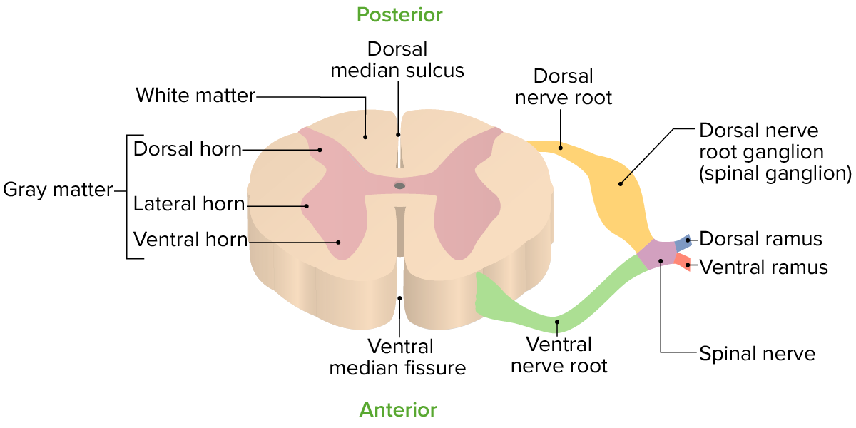
00:00 So let's talk a little bit more about cauda equina anatomy and compare conus medullaris to cauda equina syndrome. And this is very important when you're evaluating patients clinically so I want you to understand some of the key differences between these 2 syndromes. Let's start with conus medullaris syndrome. This is a spinal cord syndrome. So patients present with upper motor neuron findings like they would any other upper motor neuron spinal cord disorder. They're hyperreflexic, they have a paraparesis, and they can have bowel/bladder dysfunction and it tends to be a spastic neurogenic bladder where the bladder will just contract and compress on its own and patients have urge incontinence or urgency with their bladder dysfunction. 00:46 That's contrasted to cauda equina syndrome. Cauda equina syndrome is a peripheral nervous system disorder. This is compression of the peripheral nerves. Those peripheral nerve roots in the cauda equina of the lumbosacral region of the spinal cord and patients present with a peripheral nervous system syndrome. They can be weak, but they're hypo or areflexic. They can have urinary incontinence, but it is a urinary retention with overflow incontinence as a result of loss of tone to a flaccid bladder. 01:19 And so those are the 2 differences in those lower spinal cord syndromes that we can see; conus medullaris being a central nervous system presentation; cauda equina, a peripheral nervous system presentation and we can see the key clinical symptoms to differentiate those. The MRI is the key modality of choice for evaluating both patients and determining the appropriate management. So let's quickly compare cauda equina and conus medullaris syndromes. First, in terms of the vertebral levels that can be involved, we see similar levels that can be involved. Conus medullaris should involve the lower part of the spinal cord which typically ends around L1-2 and so that's where the pathology occurs whereas cauda equina can be from L2 all the way down to the sacral nerve roots. The spinal level that's involved, we can see sacral cord segments and roots for conus medullaris and just a lumbosacral nerve roots for cauda equina so they can look very similar in terms of their distribution. 02:17 The presentation is typically sudden and bilateral for conus medullaris. All the nerves for motor and sensory information is contained within the spinal cord in a small area and so pathology in that conus affects typically both legs and both legs bilaterally. In comparison with cauda equina, we see gradual onset of symptoms that may be asymmetric and unilateral. These different nerves are picked off depending on where the pathology develops. And that's a big difference between these 2 syndromes. Radicular pain is often less severe in conus medullaris and more severe in cauda equina as we saw in our case. Low back pain is present more often in conus medullaris and less often in cauda equina syndrome where we may have radiculopathy pain as opposed to isolated low back pain. In terms of motor strength, with conus medullaris syndrome we can see symmetric weakness that is often less marked with hyperreflexia which is really really important and we can see fasciculations as a result of loss of gray matter dysfunction at the level of the conus medullaris pathology. In terms of strength changes with cauda equina, we see that those are more markedly asymmetric. We can see areflexia, paraplegia, and atrophy is more common given the typical longer onset of that syndrome. What about reflexes? This is very important. With conus medullaris, we can see reduced ankle jerk reflexes but typically there is hyperreflexia with deep tendon reflexes in conus medullaris syndrome and in comparison, we can see areflexia or hyporeflexia with cauda equina. 03:55 In terms of sensory changes, sensory changes are localized to numbness in the perianal area. That's often symmetric and bilateral in conus medullaris and in contrast with cauda equina we can see asymmetrical sensory changes that may be unilateral. 04:10 Sphincter interrogation and sphincter dysfunction is common and must be done. 04:14 With conus medullaris, we can see early urinary and fecal incontinence and those tend to present late in cauda equina syndrome. And impotence is frequent with conus medullaris and less frequent with cauda equina. So the important table to consider when evaluating these patients or considering these questions as you evaluate patients.
About the Lecture
The lecture Myelopathy – Conus Medullaris vs. Cauda Equina by Roy Strowd, MD is from the course Diseases of the Spinal Cord.
Included Quiz Questions
Which of the following signs/symptoms would be seen with a conus medullaris lesion?
- Spastic bladder/urge incontinence
- Flaccid bladder/overflow incontinence
- Hyporeflexia
- Urinary retention
- Diarrhea
What vertebral level does the conus medullaris involve?
- L1–L2
- S2–S4
- L3–L4
- L5–S1
- T12–L1
Which reflex abnormalities are seen with cauda equina syndrome?
- Both knee and ankle reflexes decreased or absent
- Knee reflexes decreased or absent
- Both knee and ankle reflexes increased
- Brachial reflexes absent
- Flexor plantar reflexes/toes downgoing
Author of lecture Myelopathy – Conus Medullaris vs. Cauda Equina

Roy Strowd, MD
Customer reviews
5,0 of 5 stars
| 5 Stars |
|
5 |
| 4 Stars |
|
0 |
| 3 Stars |
|
0 |
| 2 Stars |
|
0 |
| 1 Star |
|
0 |
